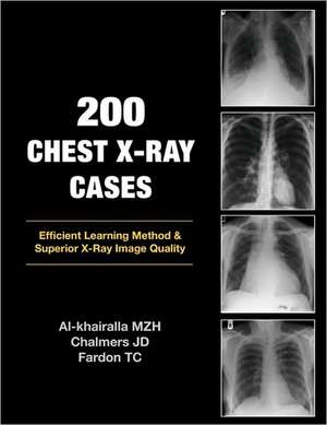
200 Chest X-Ray Cases
Autor Mudher Al-Khairalla, James Chalmers, Tom Fardonen Limba Engleză Paperback – 20 oct 2009
Preț: 358.64 lei
Preț vechi: 377.52 lei
-5% Nou
Puncte Express: 538
Preț estimativ în valută:
68.65€ • 74.59$ • 57.70£
68.65€ • 74.59$ • 57.70£
Carte tipărită la comandă
Livrare economică 21 aprilie-05 mai
Preluare comenzi: 021 569.72.76
Specificații
ISBN-13: 9781905006366
ISBN-10: 1905006365
Pagini: 216
Dimensiuni: 216 x 279 x 14 mm
Greutate: 0.51 kg
Editura: London Press
ISBN-10: 1905006365
Pagini: 216
Dimensiuni: 216 x 279 x 14 mm
Greutate: 0.51 kg
Editura: London Press
Descriere
Modern medical practice has seen many advances in imaging over the past ten years. Magnetic Resonance Imaging, CT scanning and Ultrasound investigations have all been added to the repertoire of normal practice. However, the humble chest X-ray remains a crucial first line investigation - particularly for acute medical admissions. Many chest X-rays are requested for a specific purpose e.g. confirmation of pneumonia, but the image may reveal features of previously unsuspected disease of another body system. Digital storage of X-ray images means that a chest X-ray may be viewed at any computer workstation in your hospital. Clinicians now have the opportunity to view these images without waiting for the X-ray packet to be delivered. This can only be advantageous for the patient if the clinician knows what to look for on the image. This book takes you through 200 images in a stimulating manner designed to improve your confidence in reporting the humble chest X-ray.


