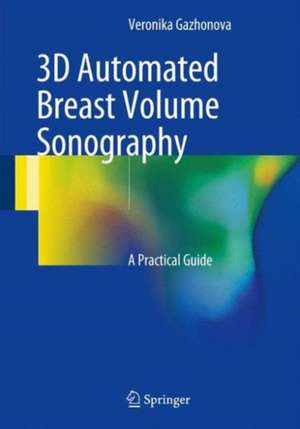
3D Automated Breast Volume Sonography: A Practical Guide
Autor Veronika Gazhonovaen Limba Engleză Hardback – 16 dec 2016
Preț: 392.60 lei
Nou
Puncte Express: 589
Preț estimativ în valută:
75.12€ • 78.65$ • 62.16£
75.12€ • 78.65$ • 62.16£
Carte disponibilă
Livrare economică 15-29 martie
Preluare comenzi: 021 569.72.76
Specificații
ISBN-13: 9783319419701
ISBN-10: 3319419706
Pagini: 327
Ilustrații: XXII, 122 p. 96 illus., 93 illus. in color.
Dimensiuni: 178 x 254 x 20 mm
Greutate: 0.52 kg
Ediția:1st ed. 2017
Editura: Springer International Publishing
Colecția Springer
Locul publicării:Cham, Switzerland
ISBN-10: 3319419706
Pagini: 327
Ilustrații: XXII, 122 p. 96 illus., 93 illus. in color.
Dimensiuni: 178 x 254 x 20 mm
Greutate: 0.52 kg
Ediția:1st ed. 2017
Editura: Springer International Publishing
Colecția Springer
Locul publicării:Cham, Switzerland
Cuprins
Introduction.- The history of the appearance and development of the automatic whole breast sonography (3D ABVS).- Current state of the 3D ABVS.- 3D ABVS Technique on ACUSON S2000 ABVS.- Clinical application of 3D ABVS for breast studies.- Normal breast.-Fibroadenoma.- Breast cystic disease.- Breast cancer.- Male breast.- Mastitis.- Implants.- Breast scar deformity.- Limitations and artifacts.- Conclusion.
Notă biografică
Veronika Gazhonova, MD, PhD, is a consultant and chief ultrasound specialist at the United Hospital and Policlinic, Moscow, Russia and Professor of Radiology and Chief Radiology Chair in the Postgraduate Medical Education & Research Center, President Medical Center, Moscow. Her areas of clinical interest are innovations in breast ultrasonography, including 3D US, sonoelastography, US-guided procedures, and contrast US. She has previously published five books as well as more than 100 articles in Russian journals and 19 publications available via ResearchGate. Dr. Gazhonova is a member of the Russian Association of Radiology (RAR), the European Society of Radiology (since 1991), and the Russian Association of Ultrasound in Medicine and Biology (RAUMB).
Textul de pe ultima copertă
This book introduces an exciting new method for breast ultrasound diagnostics – automated whole-breast volume scanning (3D ABVS). Scanning technique is described in detail, with guidance on scanning positions and protocols. Imaging findings are then illustrated and discussed for normal breast variants, the different forms of breast cancer, fibroadenomas, cystic disease, benign and malignant male breast disorders, mastitis, breast implants, and postoperative breast scars. In order to aid appreciation of the benefits of 3D ABVS, comparisons with findings on X-ray mammography and conventional 2D hand-held US are presented. Readers will be especially impressed by the convincing demonstration of the advantages of the new method for diagnosis of breast cancer in women with dense glandular tissue. In enabling readers to learn how to perform and interpret 3D ABVS, this book will be of great value for all who are embarking on its use. It will also serve as a welcome reference for radiologists, oncologists, and ultrasonographers who already have some familiarity with the technique.
Caracteristici
Introduces an exciting new method with important diagnostic advantages Describes scanning technique in detail Illustrates imaging findings across the entire range of breast pathology Presents image interpretation in comparison to X-ray mammography and hand-held ultrasonography
