Abdominal Ultrasound: Step by Step: Cărți Recomandate de SPDM
Autor Berthold Blocken Limba Engleză Paperback – 17 noi 2015
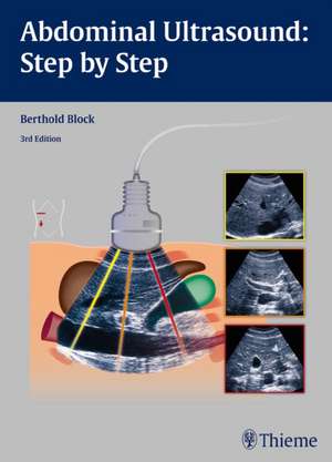
The third edition of this practical reference guide has been updated with a modern, visually attractive design and expanded content. The book is ideal for healthcare professionals with little or no experience in administering and interpreting abdominal ultrasound examinations. It is practice-oriented and structured in a way that allows readers with varying degrees of ultra-sonography knowledge to utilize the material according to their individual experience and needs.
Each chapter includes a systematic, detailed description of the anatomy involved in the ultrasound examination, with easy-to-digest steps that follow standardized routine and protocol. That straight-forward approach, coupled with more than 1,000 high-quality images and illustrations, enables hands-on learning, yielding the ability to assimilate these techniques quickly and adeptly.
This is a stellar resource that provides the requisite tools to locate and display the anatomical structure being tested, position and move the transducers accurately, describe and interpret the findings correctly, and differentiate key findings from the many image artifacts that typically occur.
Key Highlights:
- In-depth discussion of organ boundaries, organ details, anatomical relationships, potentially abnormal findings, tips, and clearly defined learning objectives
- Anatomical drawings incorporate a “sliced 3-D” view that show how the structures are displayed by the sector-shaper beam
- Each chapter includes a series of images replicating the 3-D impression that results from the transducer moving across the body
- Schematic drawings illustrate the ultrasound images, including a body marker that shows the transducer position
- The ""sono-consultant"": a systematic guide to evaluating ultrasound findings and establishing a differential diagnosis
Din seria Cărți Recomandate de SPDM
- 23%
 Preț: 877.94 lei
 - 27%
 Preț: 46.98 lei
 - 26%
 Preț: 315.93 lei
 - 5%
 Preț: 307.51 lei
 - 32%
 Preț: 47.36 lei
 - 14%
 Preț: 289.26 lei
 - 19%
 Preț: 744.33 lei
 - 19%
 Preț: 1497.54 lei
 - 19%
 Preț: 1808.18 lei
 - 19%
 Preț: 922.72 lei
 - 20%
 Preț: 2885.03 lei
 - 19%
 Preț: 508.01 lei
 - 23%
 Preț: 508.17 lei
 - 18%
 Preț: 2700.94 lei
 - 5%
 Preț: 60.73 lei
 - 23%
 Preț: 1334.67 lei
 - 19%
 Preț: 1264.50 lei
 - 24%
 Preț: 401.90 lei
 - 18%
 Preț: 2797.39 lei
 - 23%
 Preț: 1723.32 lei
 - 25%
 Preț: 2164.76 lei
 - 24%
 Preț: 1185.66 lei
 - 5%
 Preț: 4065.44 lei
 - 5%
 Preț: 2230.18 lei
 - 24%
 Preț: 601.40 lei
 - 17%
 Preț: 489.74 lei
 - 5%
 Preț: 1763.08 lei
 - 5%
 Preț: 1191.11 lei
 - 18%
 Preț: 439.75 lei
 - 5%
 Preț: 576.53 lei
 - 15%
 Preț: 268.84 lei
 - 22%
 Preț: 1456.25 lei
 - 5%
 Preț: 1308.73 lei
 - 5%
 Preț: 2762.57 lei
 - 26%
 Preț: 320.67 lei
 - 26%
 Preț: 230.11 lei
 - 23%
 Preț: 255.65 lei
 - 24%
 Preț: 188.73 lei
 - 5%
 Preț: 363.00 lei
 - 17%
 Preț: 446.36 lei
 - 31%
 Preț: 557.40 lei
 - 29%
 Preț: 434.82 lei
 - 31%
 Preț: 662.74 lei
 - 5%
 Preț: 390.20 lei
 - 23%
 Preț: 2750.17 lei
Preț: 618.53 lei
Preț vechi: 651.08 lei
-5% Nou
Puncte Express: 928
Preț estimativ în valută:
118.39€ • 128.64$ • 99.51£
118.39€ • 128.64$ • 99.51£
Carte disponibilă
Livrare economică 26 martie-01 aprilie
Livrare express 15-21 martie pentru 64.82 lei
Preluare comenzi: 021 569.72.76
Specificații
ISBN-13: 9783131383631
ISBN-10: 3131383631
Pagini: 352
Ilustrații: 1035
Dimensiuni: 199 x 277 x 26 mm
Greutate: 1.24 kg
Ediția:3rd Edition
Editura: MM – Thieme
Seria Cărți Recomandate de SPDM
ISBN-10: 3131383631
Pagini: 352
Ilustrații: 1035
Dimensiuni: 199 x 277 x 26 mm
Greutate: 1.24 kg
Ediția:3rd Edition
Editura: MM – Thieme
Seria Cărți Recomandate de SPDM
V-ar putea interesa
-
 Cope's Early Diagnosis of the Acute AbdomenWilliam Silen-16%Preț: 282.51 lei336.56 lei
 -
 Netter's Surgical Anatomy Review P.R.N.Robert B. Trelease-29%Preț: 158.51 lei224.35 lei
 -
 -15%Preț: 54.56 lei64.24 lei
 -
 -5%Preț: 284.93 lei299.92 lei
 -
 -5%Preț: 510.53 lei537.40 lei
 -
 The Family UpstairsLisa JewellPreț: 57.86 lei
 -
 The Korean Skincare BibleLeah Ganse-22%Preț: 65.61 lei84.13 lei
 -
 Top KnifeMD Hirshberg, Asher-17%Preț: 228.53 lei275.03 lei
 -
 Normal Findings in CT and MRI, A1, printTorsten Bert MöllerPreț: 161.73 lei
 -
 Preț: 343.49 lei
 -
 Der Gastroskopie-TrainerGuido Schachschal-5%Preț: 1008.79 lei1061.89 lei
 -
 Der Sono-GuideBerthold Block-5%Preț: 157.28 lei165.55 lei
 -
 Facharztprüfung Innere MedizinBerthold Block-5%Preț: 829.58 lei873.24 lei
 -
 -5%Preț: 352.93 lei371.50 lei
 -
 -15%Preț: 510.72 lei600.85 lei
 -
 Das Kalkbrennen im Schachtofen mit MischfeuerungBerthold BlockPreț: 420.20 lei
 -
 Fashion: A History from the 18th to the 20th Century:Kyoto Costume InstitutePreț: 163.95 lei
Textul de pe ultima copertă
The third edition of this practical reference guide has been updated with a modern, visually attractive design and expanded content. The book is ideal for healthcare professionals with little or no experience in administering and interpreting abdominal ultrasound examinations. It is practice-oriented and structured in a way that allows readers with varying degrees of ultra-sonography knowledge to utilize the material according to their individual experience and needs.
Each chapter includes a systematic, detailed description of the anatomy involved in the ultrasound examination, with easy-to-digest steps that follow standardized routine and protocol. That straight-forward approach, coupled with more than 1,000 high-quality images and illustrations, enables hands-on learning, yielding the ability to assimilate these techniques quickly and adeptly.
This is a stellar resource that provides the requisite tools to locate and display the anatomical structure being tested, position and move the transducers accurately, describe and interpret the findings correctly, and differentiate key findings from the many image artifacts that typically occur.
Key Highlights:
Each chapter includes a systematic, detailed description of the anatomy involved in the ultrasound examination, with easy-to-digest steps that follow standardized routine and protocol. That straight-forward approach, coupled with more than 1,000 high-quality images and illustrations, enables hands-on learning, yielding the ability to assimilate these techniques quickly and adeptly.
This is a stellar resource that provides the requisite tools to locate and display the anatomical structure being tested, position and move the transducers accurately, describe and interpret the findings correctly, and differentiate key findings from the many image artifacts that typically occur.
Key Highlights:
- In-depth discussion of organ boundaries, organ details, anatomical relationships, potentially abnormal findings, tips, and clearly defined learning objectives
- Anatomical drawings incorporate a “sliced 3-D” view that show how the structures are displayed by the sector-shaper beam
- Each chapter includes a series of images replicating the 3-D impression that results from the transducer moving across the body
- Schematic drawings illustrate the ultrasound images, including a body marker that shows the transducer position
- The ""sono-consultant"": a systematic guide to evaluating ultrasound findings and establishing a differential diagnosis
Descriere
The third edition of this practical reference guide has been updated with a modern, visually attractive design and expanded content. The book is ideal for healthcare professionals with little or no experience in administering and interpreting abdominal ultrasound examinations. It is practice-oriented and structured in a way that allows readers with varying degrees of ultra-sonography knowledge to utilize the material according to their individual experience and needs.
Each chapter includes a systematic, detailed description of the anatomy involved in the ultrasound examination, with easy-to-digest steps that follow standardized routine and protocol. That straight-forward approach, coupled with more than 1,000 high-quality images and illustrations, enables hands-on learning, yielding the ability to assimilate these techniques quickly and adeptly.
This is a stellar resource that provides the requisite tools to locate and display the anatomical structure being tested, position and move the transducers accurately, describe and interpret the findings correctly, and differentiate key findings from the many image artifacts that typically occur.
Key Highlights:
Each chapter includes a systematic, detailed description of the anatomy involved in the ultrasound examination, with easy-to-digest steps that follow standardized routine and protocol. That straight-forward approach, coupled with more than 1,000 high-quality images and illustrations, enables hands-on learning, yielding the ability to assimilate these techniques quickly and adeptly.
This is a stellar resource that provides the requisite tools to locate and display the anatomical structure being tested, position and move the transducers accurately, describe and interpret the findings correctly, and differentiate key findings from the many image artifacts that typically occur.
Key Highlights:
- In-depth discussion of organ boundaries, organ details, anatomical relationships, potentially abnormal findings, tips, and clearly defined learning objectives
- Anatomical drawings incorporate a “sliced 3-D” view that show how the structures are displayed by the sector-shaper beam
- Each chapter includes a series of images replicating the 3-D impression that results from the transducer moving across the body
- Schematic drawings illustrate the ultrasound images, including a body marker that shows the transducer position
- The ""sono-consultant"": a systematic guide to evaluating ultrasound findings and establishing a differential diagnosis