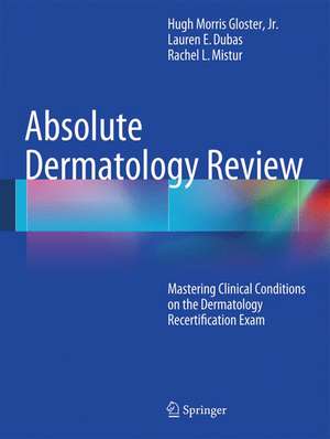
Absolute Dermatology Review: Mastering Clinical Conditions on the Dermatology Recertification Exam
Autor Hugh Morris Gloster, Jr., Lauren E. Gebauer, Rachel L. Misturen Limba Engleză Paperback – 11 iun 2015
Review of Clinical Conditions for the Dermatology Recertification Examination provides a thorough, concise review of clinical images of the specific conditions that the reader will be required to recognize during the American Board of Dermatology recertification test. In addition, concise key clinical features for each image will be provided that will assist the reader in recognizing the clinical images on the examination, enabling them a more efficient way to study for the test without having to look up images online or in a large text book. Written by a board certified dermatologic surgeon who recently took the recertification exam, this book proves indispensable to dermatologists taking the exam or residents who want a quick reference of the clinical appearances of the main conditions generally encountered by a dermatologist.
Preț: 709.11 lei
Preț vechi: 746.42 lei
-5% Nou
135.69€ • 145.10$ • 113.13£
Carte tipărită la comandă
Livrare economică 14-21 aprilie
Specificații
ISBN-10: 3319032178
Pagini: 200
Ilustrații: XVII, 534 p. 769 illus. in color.
Dimensiuni: 210 x 279 x 25 mm
Greutate: 1.23 kg
Ediția:2016
Editura: Springer International Publishing
Colecția Springer
Locul publicării:Cham, Switzerland
Public țintă
Professional/practitionerCuprins
Table of Contents
Absolute Dermatology Review
Mastering Clinical Conditions on the Dermatology Recertification Exam
Author: Hugh Gloster, Jr., MD
Section One: Papulosquamous and Eczematous Dermatoses
1. Psoriasis
a. Chronic plaque
b. Erythrodermic
c. Pustular
d. Palmoplantar
e. Guttate
f. Nail disease2. Lichen planus
a. Hypertrophic
b. Oral
c. Nails
d. Vulvovaginal
e. Annular
f. Drug induced
3. Dermatitis
a. Contact
b. Seborrheic
c. Atopic
d. Perioral
e. Stasis
f. Asteatotic
g. Dishydrotic
4. Keratosis pilaris
5. Lichen simplex chronicus
6. Lichen striatus
7. Pityriasis lichenoides
a. Pityriasis lichenoides et varioliformis acuta
b. Pityriasis lichenoides chronica
8. Pityriasis rosea
9. Pityriasis alba
10. Pityriasis rubra pilaris
11. Axillary granular parakeratosis
Section Two: Cutaneous vasculitis, vasculopathy, and microvascular occlusion syndromes
12. Small vessel (leukocytoclastic) vasculitis
a. Henoch-Schonlein purpura
b. Acute hemorrhagic edema of infancy
c. Urticarial vasculitis
d. Erythema elevatum diutinum
13. Mixed small and medium vessel vasculitis
a. Cryoglobulnemic vasculitis
b. ANCA- associated vasculitis
i. Microscopic polyangitis
ii. Wegener’s granulomatosis
iii. Churg-Strauss syndrome
14. Medium size vasculitis
a. Polyarteritis nodosa
15. Livedo vasculopathy and livedo reticularis
16. Coumadin necrosis
Section Three: Vesiculobullous diseases
17. Bullous pemphigoid
18. Cicatricial pemphigoid
19. Pemphigoid gestationis
20. Pemphigus vulgaris
21. Pemphigus foliaceous
22. Pemphigus vegetans
23. Benign familial pemphigus (Hailey-Hailey disease)
24. Dermatitis herpetiformis
25. Linear IgA bullous dermatosis
26. Epidermolysis bullosa
a. Simplex
b. Dystrophic
c. Acquired
d. Junctional
27. Newborn
a. Erythema toxicum neonatorum
28. Paraneoplastic pemphigus
Section Four: Adnexal diseases
29. Acne vulgaris
30. Rosacea
31. Fox Fordyce disease
32. Hidradenitis suppurativa
Section Five: Autoimmune disorders
33. Lupus erythematous
a. Discoid
b. Subacute cutaneous
c. Neonatal
d. Systemic
e. Acute cutaneous
f. Tumid
g. Panniculitis
h. Pernio
i. Bullous34. Dermatomyositis
35. Scleroderma and sclerodermoid skin conditions
a. Systemic sclerosis
b. Morphea
i. Plaque
ii. Linear
iii. Generalized
c. Eosinophilic fasciitits
d. Lichen sclerosus
e. Nephrogenic systemic fibrosis
f. Lipodermatosclerosis
36. Graft versus host disease
Section Six: Metabolic and deposition diseases
37. Mucinoses
a. Scleromyedema
b. Scleredema
c. Pretibial myxedema
d. Follicular mucinosis
38. Amyloidosis
39. Porphyria cutanea tarda and pseudoporphyrias
40. Calcifying disorders
a. Calcinosis cutis
b. Calciphylaxis
41. Nutritional diseases
a. Scurvy
b. Pellagra
c. Zinc deficiency (acrodermatitis enteropathica)
42. Ochronosis
Section Seven: Cutaneous manifestations of systemic diseases
43. Rheumatoid arthritis
44. Diabetes
a. Necrobiosis lipoidica
b. Acanthosis nigricans
c. Bullous diabeticorum
d. Diabetic dermopathy
e. Neuropathic ulcers
f. Scleredema diabeticorum
g. Eruptive xanthomas
45. Thyroid disease
a. Pretibial myxedema
b. Generalized myxedema
c. Acquired ichthyosis
d. Alopecia
46. Cushing’s syndrome
47. Addison’s disease
48. Gastrointestinal
a. Ulcerative colitis and Crohn’s
i. Erythema nodosum
ii. Leukocytoclastic vasculitis
iii. Sweet’s syndrome iv. Pyoderma gangrenosum
v. Acrodermatitis enteropathica
b. Gastrointestinal bleeding
i. Hereditary hemorrhagic telangiectasia
ii. Blue rubber bleb nevus syndrome
iii. Pseudoxanthoma elasticum iv. Peutz-Jeuger
v. Muir-Torre
c. Liver disease
i. Telangiectasias
ii. Spider angiomas
iii. Palmar erythema iv. Terry’s nails
v. Primary biliary cirrhosis
1. Tuberous xanthomas
vi. Hemochromatosis
vii. Wilson’s disease
49. Renal disease
a. Nephrogenic systemic sclerosis
50. Internal malignancy
a. Acanthosis nigricans
b. Acrokeratosis neoplastica (Bazek’s syndrome)
c. Erythema gyratum repens
d. Necrolytic migratory erythema
e. Acquired hypertrichosis lagunosa
f. Cutaneous metastases
g. Paraneoplastic pemphigus
Section Eight: Genodermatoses and developmental anomalies
51. Genodermatoses
a. Darier’s disease
b. Ichthyoses
i. vulgaris
ii. lamellar
iii. x-linked
iv. bullous congenital ichthyosiform erythroderma
v. Netherton’s syndrome
c. neurofibromatosis
d. tuberous sclerosus
e. Incontinentia pigmenti
f. Cowden’s syndrome
g. Cutis laxa
h. Ehlers-Danlos syndrome
i. Pachonychia congenita
j. Pseudo xanthoma elasticum
52. Developmental anomalies
a. Aplasia cutis congenita
b. Hair collar sign
Section Nine: Infections
53. Viral
a. Condyloma acuminata
b. Fifth disease
c. Hand-foot-mouth
d. Herpes simplex
i. Eczema herpeticum
ii. Herpetic Whitlow
iii. Neonatal
e. Herpes zoster
f. Varicella
g. Molluscum contagiosum
h. Verruca
54. Fungal
a. Deep fungal infections
i. Blastomycosis
ii. Sporotrichosis
iii. Coccidiodomycosis
iv. Cryptococcosis
b. Folliculitis
i. Dermatophyte
1. Tinea barbae
2. Majocchi’s
ii. Pityrosporum
c. Onychomycosis
d. Tinea
i. Pedis and manuum ii. Corporis
iii. Faceii
1. Tinea incognito
2. Tinea barbae
iv. Versicolor
v. Capitis
vi. Cruris55. Bacterial
a. Cellulitis
i. Perianal streptococcal cellulitis
b. Erythrasma
c. Folliculitis
d. Gonococcemia
e. meningococcemia
f. Impetigo
i. Non bullous ii. Bullous
g. Leishmaniasis
h. Leprosy
i. Mycobacterial infections
i. Tuberculosis
ii. Non tuberculous
j. Pitted keratolysis
k. Rocky mountain spotted fever
l. Syphilis
Section Ten: Infestations and bites
56. Cutaneous larva migrans
57. Bed bugs
58. Spider bites
59. Scabies
60. Fleas
61. Pediculosis (lice)
62. Tungiasis
Section Eleven: Hair, nails, and mucous membranes
63. Alopecias
a. Alopecia areata
b. Telogen effluvium
c. Anagen effluvium
d. Androgenetic alopecia
e. Trichotillomania
f. Scarring alopecias
i. Central centrifugal cicatricial alopecia
ii. Lichen planopilaris
iii. Discoid lupus iv. Dissecting cellulitis
v. Folliculitis decalvans
vi. Acne keloidalis nuchae
vii. Frontal fibrosing alopecia
viii. Traction alopecia
64. Nail diseasea. Trachonychia
b. Beau’s lines
c. Mee’s lines
d. Half and half nails
e. Yellow nail syndrome
f. Onychorrhexis
g. Punctate leukonychia
h. Striate leukonychia
i. Onychomadesis
j. Koilonychia
k. Terry’s nailsl. Clubbing
65. Mucous membrane disease
a. Geographic tongue
b. Oral hairy leukoplakia
c. Aphthous stomatitis
Section Twelve: Benign neoplasms
66. Accessory tragus
67. Accessory digit
68. Acquired digital fibrokeratoma
69. Adnexal tumors
a. Trichoepithelioma
b. Cylindroma
c. Syringoma
d. Nevus sebaceous
e. Trichofolliculoma
f. Poroma
70. Becker’s nevus
71. Cysts
a. Epidermal
b. Steatocystoma multiplex
c. Eruptive vellus hair cyst
72. Dermatofibroma
73. Melanocytic
a. Halo nevi
b. Congenital nevi
c. Junctional nevi
d. Compound nevi
e. Intradermal nevi
f. Nevus spilus
g. Spitz nevi
h. Blue nevi
i. Nevus of Ito
j. Nevus of Ota
k. Nevus comedonicus
74. Mucocele
75. Pyogenic granuloma
76. Seborrheic keratosis
77. Skin tags
78. Supernumerary nipples
79. Confluent and reticulated papillomatosis
80. Linear epidermal nevus
Section Thirteen: Malignant and premalignant neoplasms
81. Actinic keratosis
82. Actinic cheilitis
83. Angiosarcoma
84. Cutaneous metastases
85. Dermatofibrosarcoma protuberans
86. Kaposi’s sarcoma
87. Keratoacanthoma
88. Melanoma
89. Basal cell carcinoma
90. Squamous cell carcinoma
a. Erythroplasia of Querat
b. Verrucous carcinoma
c. Bowen’s diseased. Invasive SCC
91. Paget’s disease
92. Extramammary Paget’s disease
93. Cutaneous lymphomas
a. T-cell
i. Mycosis fungoides
ii. Folliculotrophic mycosis fungoides
iii. Pagetoid reticulosis
iv. Granulomatous slack skin
v. Sezary syndrome
vi. Primary cutaneous CD 30+ anaplastic large cell lymphoma
vii. Lymphomatoid papulosis
viii. Subacute panniculitis like T-cell lymphoma
b. B-cell
i. Follicular center lymphoma
ii. Marginal zone B-cell lymphoma
Section Fourteen: Disorders due to physical agents
94. Chondrodermatitis nodularis helicis
95. Factitial disease
96. Prurigo nodularis
97. Trauma induced skin disease
a. Talon noir
b. Erythema ab igne
c. Corns and callusesd. Subungual hematoma
e. Surgical ecchymoses
f. Piezogenic papules
g. Traumatic tattoo
98. Photodermatoses
a. Phototoxicity
b. Photoallergy
c. Actinic damage
i. Actinic purpura
ii. Poikiloderma of Civatted. Polymorphous light eruption
e. Chronic actinic dermatitis
99. Foreign body granulomas
100. Radiation dermatitis
Section Fifteen: Drug reactions, erythemas, and urticarias
101. Drug reactions
a. Acute generalized exanthematous pustulosis (AGEP)
b. Drug induced hyperpigmentation
c. Drug reaction with eosinophilia and systemic symptoms (DRESS)
d. Fixed drug eruptionse. Stevens Johnson Syndrome and Toxic epidermal necrolysis (TEN)
f. Steroid induced atrophy
g. Morbilliform
102. Erythemas
a. Erythema multiforme
b. Figurate erythemas
i. Erythema annulare centrifugum
ii. Erythema gyratum repens
iii. Erythema chronicum migransc. Necrolyitic acral erythema
d. Necrolyitic migratory erythema
103. Urticarias
a. Urticaria
b. Angioedema
104. Polymorphic eruption of pregnancy (PUPPP)
Section Sixteen: Vascular and lymphatic disorders
105. Telangiectasia
106. Hemangioma
107. Vascular malformations
108. lymphangiomas
Section Seventeen: Pigmentary disorders
109. Vitiligo
110. Postinflammatory pigment alteration
Section Eighteen: Neutrophilic dermatoses
111. Pyoderma gangrenosum
112. Sweet’s syndrome
113. Erosive pustular dermatosis of the scalp
Section Nineteen: Disorders of Langerhans cells, macrophages, and mast cells
114. Langerhans cell histiocytosis
a. Letterer-Siwe
b. Multicentric reticulohistiocytosis
115. Xanthogranuloma
a. Juvenile xanthogranuloma
b. Necrobiotic xanthogranuloma
116. Xanthomas
a. Plane
b. Eruptive
c. tuberous
117. Sarcoidosis
118. Granuloma annulare
a. Localized
b. Disseminated
119. Necrobiosis lipoidica120. Foreign body reactions
121. Mastocytosis
a. Urticaria pigmentosa
b. Solitary mastocytoma
Section Twenty: Dermal connective tissue disorders
122. Keloids
123. Hypertrophic scars
124. Perforating diseases
a. Kyrle’s disease
b. Elastosis perforans serpiginosa
c. Reactive perforating collagenosis
Section Twenty One: Panniculitis
125. Erythema nodosum
126. Lipodermatosclerosis (sclerosing panniculitis)
Recenzii
Notă biografică
Dr. Hugh Gloster is Professor of Dermatology and Director of Mohs Micrographic Surgery & Cutaneous Oncology at the University of Cincinnati Department of Dermatology.
Textul de pe ultima copertă
Each dermatologist that was board certified after 1992 is required by the American Board of Dermatology to take a recertification exam every ten years. One of the major components of the exam is to be able to identify clinical photographs of approximately 200 skin diseases, and while there are other components to the test, they vary according to subspecialty. However, everyone is required to identify the images, so several months before the exam, the American Board of Dermatology releases a list of the skin diseases that will be tested, but does not provide the images or reveal which ones will appear.
Review of Clinical Conditions for the Dermatology Recertification Examination provides a thorough, concise review of clinical images of the specific conditions that the reader will be required to recognize during the American Board of Dermatology recertification test. In addition, concise key clinical features for each image will be provided that will assist the reader in recognizing the clinical images on the examination, enabling them a more efficient way to study for the test without having to look up images online or in a large text book. Written by a board certified dermatologic surgeon who recently took the recertification exam, this book proves indispensable to dermatologists taking the exam or residents who want a quick reference of the clinical appearances of the main conditions generally encountered by a dermatologist.
Caracteristici
Includes concise key clinical features for each image that assists the reader in recognizing the clinical images on the recertification exam
Enables the reader a more efficient way of studying for the exam
Provides a quick reference of the clinical appearances of the main conditions encountered by a dermatologist
Includes hundreds of high quality photographs illustrating the main features being described in the text
Written for dermatologists taking the American Board of Dermatology recertification exam
Descriere
Each dermatologist that was board certified after 1992 is required by the American Board of Dermatology to take a recertification exam every ten years. One of the major components of the exam is to be able to identify clinical photographs of approximately 200 skin diseases, and while there are other components to the test, they vary according to subspecialty. However, everyone is required to identify the images, so several months before the exam, the American Board of Dermatology releases a list of the skin diseases that will be tested, but does not provide the images or reveal which ones will appear.
Review of Clinical Conditions for the Dermatology Recertification Examination provides a thorough, concise review of clinical images of the specific conditions that the reader will be required to recognize during the American Board of Dermatology recertification test. In addition, concise key clinical features for each image will be provided that will assist the reader in recognizing the clinical images on the examination, enabling them a more efficient way to study for the test without having to look up images online or in a large text book. Written by a board certified dermatologic surgeon who recently took the recertification exam, this book proves indispensable to dermatologists taking the exam or residents who want a quick reference of the clinical appearances of the main conditions generally encountered by a dermatologist.
