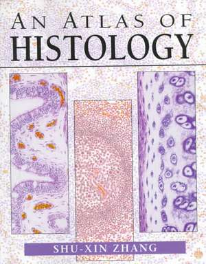
An Atlas of Histology
Autor Shu-Xin Zhangen Limba Engleză Paperback – 27 mai 1999
Preț: 791.29 lei
Preț vechi: 1029.08 lei
-23% Nou
Puncte Express: 1187
Preț estimativ în valută:
151.43€ • 157.14$ • 126.57£
151.43€ • 157.14$ • 126.57£
Carte indisponibilă temporar
Doresc să fiu notificat când acest titlu va fi disponibil:
Se trimite...
Preluare comenzi: 021 569.72.76
Specificații
ISBN-13: 9780387949543
ISBN-10: 0387949542
Pagini: 426
Ilustrații: XIX, 426 p. 393 illus.
Dimensiuni: 210 x 279 x 23 mm
Greutate: 1.35 kg
Ediția:1999
Editura: Springer
Colecția Springer
Locul publicării:New York, NY, United States
ISBN-10: 0387949542
Pagini: 426
Ilustrații: XIX, 426 p. 393 illus.
Dimensiuni: 210 x 279 x 23 mm
Greutate: 1.35 kg
Ediția:1999
Editura: Springer
Colecția Springer
Locul publicării:New York, NY, United States
Public țintă
ResearchDescriere
The beginning student of histology is frequently confronted by a paradox: diagrams in many books that illustrate human microanatomy in a simplified, cartoon-like manner are easy to understand, but are difficult to relate to actual tissue specimens or photographs. In turn, photographs often fail to show some important features of a given tissue, because no individual specimen can show all of the tissue's salient fea tures equally well. This atlas, filled with photo-realistic drawings, was prepared to help bridge the gap between the simplicity of diagrams and the more complex real ity of microstructure. All of the figures in this atlas were drawn from histological preparations used by students in my histology classes, at the level of light microscopy. Each drawing is not simply a depiction of an individual histological section, but is also a synthesis of the key structures and features seen in many preparations of similar tissues or organs. The illustrations are representative of the typical features of each tissue and organ. The atlas serves as a compendium of the basic morphological characteristics of human tissue which students should be able to recognize.
Cuprins
1. Epithelial Tissue.- 2. Connective Tissue.- 3. Cartilage and Bone.- 4. Blood Cells and Hemopoietic Cells.- 5. Muscular Tissue.- 6. Nervous Tissue and Nervous System.- 7. Circulatory System.- 8. Lymphatic Organs.- 9. Respiratory System.- 10. Digestive System.- 11. Urinary System.- 12. Male Reproductive System.- 13. Female Reproductive System.- 14. Endocrine Organs.- 15. The Integument.- 16. The Eye.- 17. The Ear.- References.
Caracteristici
Over 300 beautiful color illustrations presented in a large format
Outstanding graphical details help histology students understand complex structures
Each drawing is a compilation of the key structures and features seen in many preparations from similar tissues or organs, and doesn't simply depict an individual section
Outstanding graphical details help histology students understand complex structures
Each drawing is a compilation of the key structures and features seen in many preparations from similar tissues or organs, and doesn't simply depict an individual section
