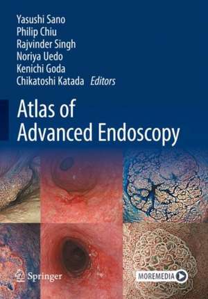
Atlas of Advanced Endoscopy
Editat de Yasushi Sano, Philip Chiu, Rajvinder Singh, Noriya Uedo, Kenichi Goda, Chikatoshi Katadaen Limba Engleză Hardback – 7 oct 2024
The book covers various aspects, including equipment reviews and diagnostic technologies using the latest unified global standard system. It explains techniques such as high-definition endoscopy, magnifying endoscopy, chromoendoscopy, NBI, TXI, RDI, and AI, while also providing insight into classifications such as the Japan Esophageal Society Classification, the VS Classification/MESDA-G, and the JNET Classification. Each case section provides expert analysis and notable endoscopic images, accompanied by histopathologic images depicting various benign lesions and early cancers.
The Atlas of Advanced Endoscopy serves as an invaluable resource to enhance the reader's knowledge and understanding of the complexities of gastrointestinal endoscopy. It is designed to help practitioners, clinicians, gastroenterologists, and medical students learn diagnostic strategies, procedures, and clinically significant cases. We hope this atlas will always be at your side in the endoscopy room.
Preț: 1113.83 lei
Preț vechi: 1172.45 lei
-5% Nou
Puncte Express: 1671
Preț estimativ în valută:
213.18€ • 230.12$ • 178.75£
213.18€ • 230.12$ • 178.75£
Carte disponibilă
Livrare economică 29 martie-12 aprilie
Preluare comenzi: 021 569.72.76
Specificații
ISBN-13: 9789819727315
ISBN-10: 9819727316
Pagini: 160
Ilustrații: Approx. 160 p. 500 illus. in color. With online files/update.
Dimensiuni: 168 x 240 mm
Greutate: 0.9 kg
Ediția:2024
Editura: Springer Nature Singapore
Colecția Springer
Locul publicării:Singapore, Singapore
ISBN-10: 9819727316
Pagini: 160
Ilustrații: Approx. 160 p. 500 illus. in color. With online files/update.
Dimensiuni: 168 x 240 mm
Greutate: 0.9 kg
Ediția:2024
Editura: Springer Nature Singapore
Colecția Springer
Locul publicării:Singapore, Singapore
Cuprins
X-1 system.- RDI.- TXI.- AI.- Paris classification.- Esophagus: The Japan esophageal society classification.- Stomach: MESDA-G.-Colon: JNET classification.- How to take high-quality images (UGI).- How to take high-quality images (LGI).- Lymphoid folicle.-LGIN/HGIN (small size).- LGIN/HGIN (large size).- Superficial cancer (flat type).- Superficial cancer (polypoid type).- Reflex esophagitis.- Papilloma.- LGIN/HGIN (small size).- LGIN/HGIN (large size).- Multple lugol-voiding lesions.- Superficial cancer (0-Ⅰ, M).- Superficial cancer (0-Ⅱ,M).- Superficial cancer (SM).- Superficial cancer (SM).- Superficiail cancer (large, ≧ 2/3 circumference).- SSBE and LSBE.- Barrett's dysplasia (small size).- Barrett's dysplasia (large size).- Barrett's adenocarcinoma (flat type).- Barrett's adenocarcinoma (polypoid type).- Ectopic gastric metaplasia.- denoma (Papillary resion).- Adenoma (Gastric type, non-papillary resion).- Adenoma (Intestinal type, non-papillary resion).- Duodenal early cancer (non-papillary resion).- Gastric metaplasia.- Hyperplastic polyp.- fundic glnad polyp.- Adenoma.- Early garstric cancer (0-Ⅰ, M, Differentiated type).- Early garstric cancer (0-Ⅱa, M, Differentiated type).- Early garstric cancer (0-Ⅱc, M, Differentiated type).- Early garstric cancer (any, SM, Differentiatted type).- Early garstric cancer (0-Ⅱc, M, Undifferentiated type).- Early garstric cancer (any, SM, Undifferentiatted type).- Early gastric cancer (UL+).- Early gastric cancer after HP eradication (1).- Early gastric cancer after HP eradication (2).- Gastric adenocarcinoma of fundic-gland type.- Gastric MALT Lymphoma.- Hyperplastic polyp (HP).- sessile serrated lesion (SSL).- SSL with dsyplasia or cancer.- Superficial serrated adenoma (SuSA).- Tradiational serrated adenoma.- Polypoid-type adenoma (0-Ⅰ).- Flat-type adenoma.- Depressed-type adenoma (0-Ⅱc).- Early colorectal cancer (0-Ⅰ, Polpyoid, M or SM).- Early colorectal cancer (0-ⅠP+Ⅱc ,Head or Stalk invasion).- Early colorectal cancer (0-Ⅱa, LST-NG, M or SM).- Early colorectal cancer (0-Ⅱa+Ⅰs, LST-G-Mix).- Early colorectal cancer (0-Ⅱa+Ⅱc, SM).- Early colorectal cancer (0-Ⅰs+Ⅱc, SM).- Anal condyloma or cancer.
Notă biografică
Prof. Yasushi Sano (FJGES) is a Clinical Professor at Kansai Medical University, as well as the Director of Gastrointestinal Center at Sano Hospital, Kobe, Japan. He is one of world leaders in the field of diagnostic and therapeutic colonoscopy. He showed many lectures regarding image enhanced endoscopy (IEE) technique and demonstrated endoscopy live with EMR/ESD technique at several countries. He skillfully uses magnifying colonoscopy with advanced imaging technique. He has published more than 180 peer reviewed manuscripts and 9 book chapters. He is a member of several advisory boards including Gastroenterological Endoscopy (JGES, JGA, Japan), Gastrointestinal Endoscopy (GIE, USA) and a chairman of Asian Novel Bio-Imaging and Intervention Group (ANBIIG). Workshops having participated in more than 230 workshops/symposiums across various countries.
Prof. Philip Chiu is Professor of Division of Upper GI and Metabolic Surgery, Department of Surgery, Director of Multi-Scale Medical Robotics Center, Director of Endoscopy Center, Institute of Digestive Disease; Director of CUHK Chow Yuk Ho Technology Center for Innovative Medicine and Dean (External Affairs), Faculty of Medicine, Chinese University of Hong Kong. Prof. Chiu is first to perform ESD for treatment of early GI cancers in Hong Kong in 2004. In 2010, he performed first Per-oral Endoscopic Myotomy (P.O.E.M.) in Hong Kong as well as pioneering world first robotic gastric ESD in 2011, followed by world first robotic colorectal ESD in 2020. His research interests include esophageal cancer management, minimally invasive and robotic esophagectomy, novel endoscopic technologies for diagnosis of early GI cancers, endoscopic surgery as well as robotics for endoluminal surgery. He has published more than 370 peer reviewed manuscripts and 6 book chapters. He is currently co-editor of Endoscopy and subject editor for Surgical Endoscopy.
Prof. Rajvinder Singh is a Professor of Medicine with the University of Adelaide and the Director of Gastroenterology at the Lyell McEwin & Modbury Hospitals, South Australia. He is an Editorial Board member of Clinical Endoscopy and past Editorial Board member of Endoscopy, Digestive Endoscopy and Endoscopy International Open. He has a h-index of 41. He has authored more than 150 publications and book chapters on Advanced Endoscopic Mucosal Imaging and Resection techniques. He has been involved in various committees drafting national and international guidelines on this subject. He was the past chair of the Australian Gastrointestinal Endoscopic Association (AGEA). He is frequently invited to conduct Basic and Advanced Endoscopy Workshops having participated in more than 200 workshops/symposiums across various countries.
Dr. Noriya Uedo is a vice-director, Department of Gastrointestinal Oncology, Osaka International Cancer Institute. He graduated from the School of Medicine, Kagoshima University in 1992 and started his training in gastrointestinal oncology and endoscopy in the current institution from 1994. He is a councilor of several Japanese medical societies. He is serving as a visiting professor in Fukuoka University Chikushi Hospital, and used to serve as a visiting professor in Lund University Malmoe University hospital, and Chinese PLA general hospital.
Prof. Kenichi Goda graduated from National Defense Medical College. He is a Gastroenterologist specialized in gastrointestinal endoscopy and the Director of Gastrointestinal Endoscopy Center of Dokkyo Medical University Hospital. He has more than 90 peer reviewed international publications and have delivered numerous oral and posters presentations in numerous international meetings. He received awards of Distinguished Paper Award of Japan GastroenterologicalEndoscopy Society 2004 and Distinguished Presentation of JDDW 2012.
Dr. Chikatoshi Katada graduated from Kitasato University School of Medicine in 1998. From 2001 to 2005, he worked at National Cancer Center Hospital East. From 2005 to 2022, he worked at Kitasato University Hospital. From 2018 to 2019, he participated in the visiting clinician program at Mayo Clinic. From 2022 to the present, he worked at Kyoto University Hospital. He has more than 120 peer reviewed international publications and has delivered numerous presentations in international meetings. His area of expertise is clinical research on carcinogenesis and prevention of alcohol-related cancers of the esophagus and head and neck. Therefore, he is well versed in the endoscopic diagnosis and treatment of cancers arising in this area.
Textul de pe ultima copertă
This comprehensive book features high-quality images, videos, and step-by-step instructions for capturing detailed views of the upper and lower gastrointestinal tract from the oral cavity to the anus. In addition to covering areas such as the tongue, head and neck, non-Barrett's esophagus, Barrett's esophagus, stomach, duodenum, colon, and rectum, the atlas is authored by leading GI experts from the Asian Novel Bio-Imaging and Intervention Group in the Asia-Pacific region, known for their unparalleled expertise and commitment to excellence.
The book covers various aspects, including equipment reviews and diagnostic technologies using the latest unified global standard system. It explains techniques such as high-definition endoscopy, magnifying endoscopy, chromoendoscopy, NBI, TXI, RDI, and AI, while also providing insight into classifications such as the Japan Esophageal Society Classification, the VS Classification/MESDA-G, and the JNET Classification. Each case section provides expert analysis and notable endoscopic images, accompanied by histopathologic images depicting various benign lesions and early cancers.
The Atlas of Advanced Endoscopy serves as an invaluable resource to enhance the reader's knowledge and understanding of the complexities of gastrointestinal endoscopy. It is designed to help practitioners, clinicians, gastroenterologists, and medical students learn diagnostic strategies, procedures, and clinically significant cases. We hope this atlas will always be at your side in the endoscopy room.
Caracteristici
Covers varieties of endoscopic images and video clips of the gastrointestinal tract Provides a detailed description and analysis of different classification systems used for various GI conditions Provides detailed information for different conditions to help clinical decision-making and treatment planning
