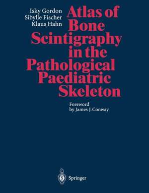
Atlas of Bone Scintigraphy in the Pathological Paediatric Skeleton: Under the Auspices of the Paediatric Committee of the European Association of Nuclear Medicine
Autor Isky Gordon Cuvânt înainte de J.J. Conway Autor Sibylle Fischer, Klaus Hahnen Limba Engleză Paperback – 16 noi 2013
Preț: 732.56 lei
Preț vechi: 771.12 lei
-5% Nou
Puncte Express: 1099
Preț estimativ în valută:
140.18€ • 146.72$ • 116.67£
140.18€ • 146.72$ • 116.67£
Carte tipărită la comandă
Livrare economică 31 martie-14 aprilie
Preluare comenzi: 021 569.72.76
Specificații
ISBN-13: 9783642646751
ISBN-10: 3642646751
Pagini: 360
Ilustrații: XI, 343 p. 461 illus.
Dimensiuni: 210 x 279 x 25 mm
Greutate: 0.84 kg
Ediția:Softcover reprint of the original 1st ed. 1996
Editura: Springer Berlin, Heidelberg
Colecția Springer
Locul publicării:Berlin, Heidelberg, Germany
ISBN-10: 3642646751
Pagini: 360
Ilustrații: XI, 343 p. 461 illus.
Dimensiuni: 210 x 279 x 25 mm
Greutate: 0.84 kg
Ediția:Softcover reprint of the original 1st ed. 1996
Editura: Springer Berlin, Heidelberg
Colecția Springer
Locul publicării:Berlin, Heidelberg, Germany
Public țintă
Professional/practitionerCuprins
1: Introduction.- 2: Infection.- 2.1 Typical Hot Lesion in Bone.- 2.2 Less Common Appearances.- 2.3 Unusual Sites, Excluding the Long Bones.- 2.4 Non-skeletal Infection.- 2.5 Growth Arrest.- 3: Arthritis.- 3.1 Septic Arthritis.- 3.2 Aseptic Arthritis.- 4: Tumours.- 4.1 Benign Tumours.- 4.2 Malignant Tumours.- 4.3 Tumour Secondaries.- 4.4 Langerhans’ Histiocytosis.- 5: Trauma.- 5.1 Appearances at Common Sites.- 5.2 Unusual Appearances or Locations of Fractures.- 5.3 Diffuse Skeletal Involvement.- 5.4 Complication of Trauma.- 5.5 Bone Response to Underlying Pathology.- 5.6 Post-operative Appearances.- 5.7 Effect of Radiotherapy.- 6: Osteochondritis Dissecans — Avascular Necrosis.- 6.1 Legg-Perthes’ Disease.- 6.2 Other Sites of Osteochondritis Dissecans.- 6.3 Sickle Cell Disease.- 7: Miscellaneous.- 7.1 Dysplasia.- 7.2 Chondromata.- 7.3 Blount’s Disease.- 7.4 Gaucher’s Disease.- 7.5 Scoliosis.- 7.6 The Sick Child.- 7.7 Disuse Arthropathy.- 7.8 Muscular Disorders.- 7.9 Growth Arrest.- 8: Unusual Appearances of the Bone-Seeking Tracer.- 8.1 Kidney and Collecting System.- 8.2 Lung Uptake.- 8.3 Splenic Uptake.- 8.4 Brain Uptake.- 8.5 Soft Tissue Calcification.- 8.6 Isotope Artefact.
Textul de pe ultima copertă
This atlas covers both the common pathologies affecting the paediatric skeleton as well as unusual pathology. Variations in the appearances of osteomyelitis have been extensively illustrated, including usual and unusual bone involvement. The images illustrated include whole body scanning, gamma camera high resolution spot images, pin holw images and SPECT. Three phase bone scans are also illustrated. Indications for the use of each type of bone scan is covered in the text. The majority of the cases illustrated are recomparied by either a Teaching Point or a Technical Comment. There is extensive cross reference pointing the similar appearances of different pathologies as seen on radioisotope bone scintigraphy. a comprehensive subject index makes it easier to find special items in the book. Extensive coverage of the Tc99m bone seeking tracer in non osseos sites is illustrated. Whilest this covers mainly the kidney, other sites are included. The atlas will allow the paediatrician, the orthopaedic surgeon, the radiologist and the nuclear medicine physician to compare the radioisotope bone scan of his/her patient with illustrations in this atlas, there increasing the physicians confidence of the final diagnosis.


