Atlas of Cell Organelles Fluorescence
Autor Elli Kohen, Rene Santus, Joseph G. Hirschberg, Nuri Ozkutuken Limba Engleză Hardback – 29 dec 2003
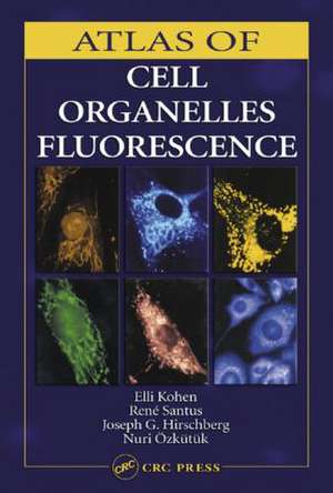
The book, an exhaustive list of cellular events observed using microfluorometry, supplies a compilation of information spread throughout the literature of the last 30 years. Atlas of Cell Organelles Fluorescence brings the information together and puts it in an easily accessible format.
Preț: 1362.53 lei
Preț vechi: 1826.80 lei
-25% Nou
Puncte Express: 2044
Preț estimativ în valută:
260.74€ • 270.68$ • 217.42£
260.74€ • 270.68$ • 217.42£
Comandă specială
Livrare economică 04-18 martie
Doresc să fiu notificat când acest titlu va fi disponibil:
Se trimite...
Preluare comenzi: 021 569.72.76
Specificații
ISBN-13: 9780849314407
ISBN-10: 0849314402
Pagini: 208
Ilustrații: 188 b/w images, 48 color images and 172 halftones
Dimensiuni: 178 x 254 x 16 mm
Greutate: 1.22 kg
Ediția:New.
Editura: CRC Press
Colecția CRC Press
Locul publicării:Boca Raton, United States
ISBN-10: 0849314402
Pagini: 208
Ilustrații: 188 b/w images, 48 color images and 172 halftones
Dimensiuni: 178 x 254 x 16 mm
Greutate: 1.22 kg
Ediția:New.
Editura: CRC Press
Colecția CRC Press
Locul publicării:Boca Raton, United States
Public țintă
ProfessionalCuprins
Vital Fluorescence Probes of Cell Organelles. Metabolic Probes. Cytotoxic Drugs. Genetic Diseases. Cell Differentiation and Cell Pathology. Cell-to-Cell Communication. The Study of Microecosystems. Biotechnology. Instrumentation. Novel Methods and Instrumental Designs. Conclusion. Index.
Notă biografică
Kohen\, Elli; Santus\, Rene; Hirschberg\, Joseph G.; Ozkutuk\, Nuri
Descriere
Containing over 150 original photomicrographs accompanied by protocol information, this book shows organelles’ structures, interaction, and organization into complexes. It includes a color insert that illustrates some of the most characteristic organelle features, microcompartmentalization, and alterations as investigated by one or more probes simultaneously. Each section provides a brief introduction and technical details of staining methods and gives explanations of unusual appearances and the cytochemical reactions when necessary. The book serves as a guide for therapeutic, diagnostic, or prognostic interpretation of images and for further research.
