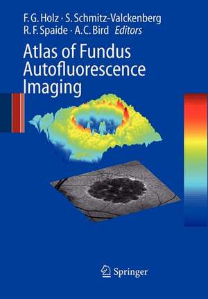
Atlas of Fundus Autofluorescence Imaging
Editat de Frank G. Holz, Steffen Schmitz-Valckenberg, Richard F. Spaide, Alan C. Birden Limba Engleză Paperback – 15 oct 2010
This unique atlas provides a comprehensive and up-to-date overview of FAF imaging in retinal diseases. It also compares FAF findings with other imaging techniques such as
fundus photograph, fluorescein- and ICG angiography as well as optical coherence tomography.
General ophthalmologists as well as retina specialists will find this a very useful guide which illustrates typical FAF characteristics of various retinal diseases.
Preț: 968.07 lei
Preț vechi: 1019.01 lei
-5% Nou
Puncte Express: 1452
Preț estimativ în valută:
185.26€ • 201.17$ • 155.62£
185.26€ • 201.17$ • 155.62£
Carte tipărită la comandă
Livrare economică 22 aprilie-06 mai
Preluare comenzi: 021 569.72.76
Specificații
ISBN-13: 9783642091193
ISBN-10: 3642091199
Pagini: 360
Ilustrații: XIII, 341 p.
Dimensiuni: 168 x 240 x 19 mm
Greutate: 0.57 kg
Ediția:Softcover reprint of hardcover 1st ed. 2007
Editura: Springer Berlin, Heidelberg
Colecția Springer
Locul publicării:Berlin, Heidelberg, Germany
ISBN-10: 3642091199
Pagini: 360
Ilustrații: XIII, 341 p.
Dimensiuni: 168 x 240 x 19 mm
Greutate: 0.57 kg
Ediția:Softcover reprint of hardcover 1st ed. 2007
Editura: Springer Berlin, Heidelberg
Colecția Springer
Locul publicării:Berlin, Heidelberg, Germany
Public țintă
ResearchCuprins
Methodology.- Lipofuscin of the Retinal Pigment Epithelium.- Origin of Fundus Autofluorescence.- Fundus Autofluorescence Imaging with the Confocal Scanning Laser Ophthalmoscope.- How To Obtain the Optimal Fundus Autofluorescence Image with the Confocal Scanning Laser Ophthalmoscope.- Autofluorescence Imaging with the Fundus Camera.- Macular Pigment Measurement—Theoretical Background.- Macular Pigment Measurement —Clinical Applications.- Evaluation of Fundus Autofluorescence Images.- Clinical Application.- Macular and Retinal Dystrophies.- Discrete Lines of Increased Fundus Autofluorescence in Various Forms of Retinal Dystrophies.- Age-Related Macular Degeneration I—Early Manifestation.- Age-Related Macular Degeneration II—Geographic Atrophy.- Age-Related Macular Degeneration III—Pigment Epithelium Detachment.- Age-Related Macular Degeneration IV—Choroidal Neovascularization (CNV).- Idiopathic Macular Telangiectasia.- Chorioretinal Inflammatory Disorders.- Autofluorescence from the Outer Retina and Subretinal Space.- Miscellaneous.- Perspectives in Imaging Technologies.- Perspectives in Imaging Technologies.
Recenzii
From the reviews:
"This book … gives readers a comprehensive overview of the physiological and anatomic changes leading to fundus autofluorescence. … The book is enriched with hundreds of high quality illustrations and fundus autofluorescence pictures as well as color pictures, fluorescein angiography and some microperimetry. The table of contents is comprehensive and clear. Each chapter is followed by a large and updated list of references. … We can thoroughly recommend this book to general ophthalmologists as well as retina specialists." (Michaela Goldstein and Anat Loewenstein, Grafe’s Archive for Clinical and Experimental Ophthalmology, Vol. 247, 2009)
"This book … gives readers a comprehensive overview of the physiological and anatomic changes leading to fundus autofluorescence. … The book is enriched with hundreds of high quality illustrations and fundus autofluorescence pictures as well as color pictures, fluorescein angiography and some microperimetry. The table of contents is comprehensive and clear. Each chapter is followed by a large and updated list of references. … We can thoroughly recommend this book to general ophthalmologists as well as retina specialists." (Michaela Goldstein and Anat Loewenstein, Grafe’s Archive for Clinical and Experimental Ophthalmology, Vol. 247, 2009)
Caracteristici
Fundus autofluorescence (FAF) imaging provides new information on diagnostics and novel therapies regarding retinal diseases Provides a comprehensive and up-to-date overview of FAF imaging in retinal diseases Lavishly illustrated atlas Written by renowned international experts Includes supplementary material: sn.pub/extras
