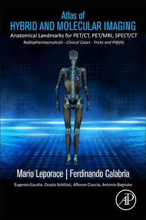
Atlas of Hybrid Imaging Sectional Anatomy for PET/CT, PET/MRI and SPECT/CT Vol. 1: Brain and Neck: Sectional Anatomy for PET/CT, PET/MRI and SPECT/CT
Autor Mario Leporace, Ferdinando Calabria, Eugenio Gaudio, Orazio Schillaci, Alfonso Ciaccio, Antonio Bagnatoen Limba Engleză Paperback – 24 mai 2023
The structure in three volumes represents an user friendly guide for interpreting PET and SPECT in relation to co-registered CT and/or MRI.
Three companion volumes with a practical structure in two-page unit offer to the reader a navigational tool, based on anatomical districts, with labeled and explained low-dose multiplanar CT or MRI views merged with PET fusion imaging on the right hand and contrast enhanced CT or MRI on the other side. This new format enables rapid identification of hybrid nuclear medicine findings which are now routine at leading medical centers.
Volume 1 is focused on brain and neck PET imaging, with emphasis on PET/MRI; Volume 2 concerns thorax, abdomen and pelvis, with particular attention on lung and liver segmental anatomy and evaluation of peritoneum. Special chapters on heart, lymph nodes and musculoskeletal system, are collected in the Volume 3.
Each chapter begins with three-dimensional CT and/or MRI views of the evaluated anatomical region, bringing forward sectional tables.
Clinical cases, tricks and pitfalls linked to several PET or SPECT radiopharmaceuticals help to introduce the reader to peculiar molecular pathways and to improve confidence in cross-sectional imaging, that is vital for the accurate diagnosis and treatment of diseases.
- Compact, comprehensive, easy to read guide to sectional imaging and multiplanar evaluation of hybrid PET and SPECT
- Includes 856 fully colored, labeled, high quality original images of axial, coronal and sagittal CT, contrast enhanced CT, PET/CT and/or PET/MRI
- Displays 200 clinical cases, showcasing both common and unusual findings that nuclear physicians and radiologists could encounter in their clinical practice
- Costant orthogonal references on 3D images allow orientation on axial, coronal and sagittal views
- Peculiar aspects of 18 PET and SPECT radiopharmaceuticals, defined by distinctive color scales
- Terms and descriptions mostly based on Terminologia Anatomica
- Specific text boxes explain anatomical variants, radiological advices and physiological findings linked to tracer bio-distribution
- Supports interpretation and report of hybrid nuclear medicine scans
- For professionals and vocationals
Preț: 740.52 lei
Preț vechi: 969.09 lei
-24% Nou
Puncte Express: 1111
Preț estimativ în valută:
141.69€ • 147.94$ • 117.27£
141.69€ • 147.94$ • 117.27£
Carte disponibilă
Livrare economică 07-21 martie
Livrare express 28 februarie-06 martie pentru 88.60 lei
Preluare comenzi: 021 569.72.76
Specificații
ISBN-13: 9780323904544
ISBN-10: 0323904548
Pagini: 196
Ilustrații: 500 illustrations (250 in full color)
Dimensiuni: 216 x 276 x 16 mm
Greutate: 0.56 kg
Editura: ELSEVIER SCIENCE
ISBN-10: 0323904548
Pagini: 196
Ilustrații: 500 illustrations (250 in full color)
Dimensiuni: 216 x 276 x 16 mm
Greutate: 0.56 kg
Editura: ELSEVIER SCIENCE
Cuprins
1. BRAIN
2. NECK and MAXILLOFACIAL REGION
2. NECK and MAXILLOFACIAL REGION
