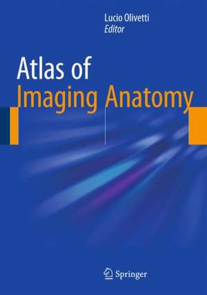
Atlas of Imaging Anatomy
Editat de Lucio Olivettien Limba Engleză Hardback – 15 ian 2015
Preț: 1298.35 lei
Preț vechi: 1366.68 lei
-5% Nou
Puncte Express: 1948
Preț estimativ în valută:
248.43€ • 260.32$ • 205.38£
248.43€ • 260.32$ • 205.38£
Carte disponibilă
Livrare economică 21 martie-04 aprilie
Preluare comenzi: 021 569.72.76
Specificații
ISBN-13: 9783319107493
ISBN-10: 3319107496
Pagini: 250
Ilustrații: XI, 274 p. 320 illus., 127 illus. in color.
Dimensiuni: 178 x 254 x 20 mm
Greutate: 0.79 kg
Ediția:2015
Editura: Springer International Publishing
Colecția Springer
Locul publicării:Cham, Switzerland
ISBN-10: 3319107496
Pagini: 250
Ilustrații: XI, 274 p. 320 illus., 127 illus. in color.
Dimensiuni: 178 x 254 x 20 mm
Greutate: 0.79 kg
Ediția:2015
Editura: Springer International Publishing
Colecția Springer
Locul publicării:Cham, Switzerland
Public țintă
Professional/practitionerCuprins
Brain.- Spine.- Head and neck.- Breast.- Chest.- Mediastinum and heart.- Abdominal cavity, peritoneum, and retroperitoneum.- Gastrointestinal tract.- Liver, biliary tract, and pancreas.- Urinary tract.- Male pelvis.- Female pelvis.- Joints.
Textul de pe ultima copertă
This book is designed to meet the needs of radiologists and radiographers by clearly depicting the anatomy that is generally visible on imaging studies. It presents the normal appearances on the most frequently used imaging techniques, including conventional radiology, ultrasound, computed tomography, and magnetic resonance imaging. Similarly, all relevant body regions are covered: brain, spine, head and neck, chest, mediastinum and heart, abdomen, gastrointestinal tract, liver, biliary tract, pancreas, urinary tract, and musculoskeletal system. The text accompanying the images describes the normal anatomy in a straightforward way and provides the medical information required in order to understand why we see what we see on diagnostic images. Helpful correlative anatomic illustrations in color have been created by a team of medical illustrators to further facilitate understanding.
Caracteristici
Depicts the normal anatomy as seen on imaging studies Explains why we see what we see on diagnostic images Covers a wide range of imaging modalities and anatomic regions Includes correlative anatomic illustrations Includes supplementary material: sn.pub/extras
