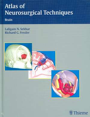
Atlas of Neurosurgical Techniques: Brain
Editat de Laligam N. Sekhar, Richard Glenn Fessleren Limba Engleză Hardback – 22 aug 2006
Atlas of Neurosurgical Techniques: Brain presents the current information on how to manage diseases and disorders of the brain.
Ideal as a reference for review in preparation for surgery, this atlas features succinct discussion of pathology and etiology that helps the reader gain a firm understanding of the underlying disease and conditions. The authors provide step-by-step descriptions of surgical techniques, clearly delineating the indications and contraindications, the goals, the operative preparation and anesthesia, and postoperative management. Common complications of techniques are also emphasized. Over 900 illustrations aid the rapid comprehension of the surgical procedures described in the text.
Highlights:
- Clear descriptions of the surgical management of aneurysms, arteriovenous malformations, occlusive and hemorrhagic vascular diseases, tumors, lesions, pain disorders, trauma, infections, and more
- Detailed discussion of disease pathology, etiology, and differential diagnosis
- Concise outlines of indications, contraindications, as well as advantages and disadvantages of each technique illuminate the rationale behind surgical management
- More than 900 illustrations, including 684 in full-color, demonstrate key concepts
- Sections on the latest techniques in stereotactic and minimally invasive surgery
Preț: 2615.05 lei
Preț vechi: 2752.68 lei
-5% Nou
Puncte Express: 3923
Preț estimativ în valută:
501.02€ • 527.47$ • 413.54£
501.02€ • 527.47$ • 413.54£
Carte indisponibilă temporar
Doresc să fiu notificat când acest titlu va fi disponibil:
Se trimite...
Preluare comenzi: 021 569.72.76
Specificații
ISBN-13: 9780865779204
ISBN-10: 0865779201
Pagini: 1104
Ilustrații: 1345
Dimensiuni: 216 x 279 x 51 mm
Greutate: 3.49 kg
Ediția:1st Edition
Editura: Thieme
Colecția Thieme
ISBN-10: 0865779201
Pagini: 1104
Ilustrații: 1345
Dimensiuni: 216 x 279 x 51 mm
Greutate: 3.49 kg
Ediția:1st Edition
Editura: Thieme
Colecția Thieme
Recenzii
Written by the 'Who's Who' of neurosurgery. The very first chapter...by Dr. Albert L. Rhoton Jr. is a must-read... The real power of this atlas lies in the clarity and simplicity of its illustrations and procedural descriptions. On this account, the book is excellent. Each chapter is richly illustrated with detailed yet simple graphics that communicate the salient issues of each procedure. [Illustrators] Jennifer Pryll and Tony Pazos...are to be commended for their high-quality work and the impact of their contributions throughout the text. ...A typical Thieme textbook with the standard dark blue and silver cover graphic...of high-quality..this text is a good value and should be strongly considered as a meaningful addition to one's collection of basic neurosurgical textbooks.--Journal of NeurosurgeryWritten by authoritative educators in their respective fields to produce an exemplary operative brain atlas...very well organized...The individual chapters are concise yet highly informative. The organization of each chapter allows for a quick review on relevant anatomy, anesthetic considerations, surgical indications, surgical technique, complications, and postoperative care, with a succinct conclusion. The sections on surgical indication give a detailed step-by-step approach, highlighting pitfalls and also management of intraoperative complications. The illustrations are straightforward and easy to follow without sacrificing useful detail...This book is a must for any neurosurgical library.--Otology & NeurotologyA comprehensive review of the topic...with superb drawings and graphs and references...a superb piece of work...This is an excellent source of information...a must in any neurosurgical library.--Doody's ReviewVolume 1: Brain and Volume 2: Spine, lays out in artist's color drawings, line diagrams, intraoperative photographs, radiographs, and in an easily digestible written format the breadth and scope of virtually every neurosurgical procedure...The illustrations...are highly educational and allow nonsurgeons to understand the approaches to various lesions and the potential pitfalls one many encounter in the operating room..Imaging is abundant...both volumes can help fill a need for practicing neuroradiology and in training programs in neuroradiology.--American Journal of Neuroradiology
Notă biografică
Neurosurgeon, Rush University Medical Center, Chicago, ILProfessor, Dept of Neurological Surgery, Northwestern University, Feinberg School of Medicine, Chicaco, IL, USA,
Textul de pe ultima copertă
Atlas of Neurosurgical Techniques: Brain presents the current information on how to manage diseases and disorders of the brain.
Ideal as a reference for review in preparation for surgery, this atlas features succinct discussion of pathology and etiology that helps the reader gain a firm understanding of the underlying disease and conditions. The authors provide step-by-step descriptions of surgical techniques, clearly delineating the indications and contraindications, the goals, the operative preparation and anesthesia, and postoperative management. Common complications of techniques are also emphasized. Over 900 illustrations aid the rapid comprehension of the surgical procedures described in the text.
Highlights:
Ideal as a reference for review in preparation for surgery, this atlas features succinct discussion of pathology and etiology that helps the reader gain a firm understanding of the underlying disease and conditions. The authors provide step-by-step descriptions of surgical techniques, clearly delineating the indications and contraindications, the goals, the operative preparation and anesthesia, and postoperative management. Common complications of techniques are also emphasized. Over 900 illustrations aid the rapid comprehension of the surgical procedures described in the text.
Highlights:
- Clear descriptions of the surgical management of aneurysms, arteriovenous malformations, occlusive and hemorrhagic vascular diseases, tumors, lesions, pain disorders, trauma, infections, and more
- Detailed discussion of disease pathology, etiology, and differential diagnosis
- Concise outlines of indications, contraindications, as well as advantages and disadvantages of each technique illuminate the rationale behind surgical management
- More than 900 illustrations, including 684 in full-color, demonstrate key concepts
- Sections on the latest techniques in stereotactic and minimally invasive surgery
Descriere
Atlas of Neurosurgical Techniques: Brain presents the current information on how to manage diseases and disorders of the brain. Ideal as a reference for review in preparation for surgery, this atlas features succinct discussion of pathology and etiology and step-by-step descriptions of surgical techniques.
Cuprins
Volume 1
Section I General Principles and Basic Techniques
1 General Techniques of Cranial Exposure
2 General Principles of Microsurgery
3 Anesthesia Techniques and Principles of Hemostasis and Blood Replacement for Cranial Surgery
4 Principles of Blood Coagulation and Transfusion
5 Neurophysiological Monitoring During Neurosurgery: Indicated Uses and Practical Considerations
6 Critical Care for Neurosurgery
7 Endovascular Surgery: General Technique
8 Endoscopic Surgery: General Principles and Transsphenoidal Approaches
8 Appendix: Endoscopic and Endoscope-Assisted Approaches to the Sellar, Suprasellar, and Ventricular Regions (Supplemental Videos)
Section II Aneurysms
9 General Principles of Aneurysm Surgery
10 Internal Carotid Artery: Supraclinoid Aneurysms
11 Internal Carotid Artery: Infraclinoid/Clinoid Aneurysms
12 Middle Cerebral Artery Aneurysms
13 Anterior Communicating Artery Aneurysms
14 Interhemispheric Approach to Anterior Communicating Artery Aneurysms
15 Distal Anterior Cerebral Artery Aneurysms
16 Distal Middle Cerebral Artery Aneurysms
17 Basilar Artery Tip and Superior Cerebellar Aneurysms
18 Midbasilar and Vertebrobasilar Junction Aneurysms: Extended Retrosigmoid Approach
19 Vertebral Artery and Posterior Inferior Cerebellar Artery Aneurysms
20 Cranial Base Approaches to Aneurysms
21 Microsurgery of Giant Intracranial Aneurysms
22 Giant Aneurysms
23 Endovascular Coiling of Aneurysms
23 Appendix: Endovascular Complication Management (Supplemental Videos)
24 Stent-Assisted Coiling and Flow Diversion for Intracranial Aneurysms
25 Cerebral Revascularization for Aneurysms and Skull Base Tumors
26 In Situ Bypasses for Intracranial Aneurysms
Section III Arteriovenous Malformations
27 General Techniques for the Surgery of Arteriovenous Malformations
28 Preoperative and Therapeutic Embolization of Arteriovenous Malformation with N-Butyl-2-cyanoacrylate
29 Embolization of Arteriovenous Malformations with Onyx and Combined Treatments
30 Frontal, Occipital, and Temporal Arteriovenous Malformations
31 Perimotor and Perisylvian Arteriovenous Malformations
32 Corpus Callosum and Deep Arteriovenous Malformations
33 Posterior Fossa Arteriovenous Malformations
34 Cavernous Malformations of the Brain
35 Brainstem Cavernous Malformations
36 Carotid-Cavernous Fistula
37 Vein of Galen Malformations
38 Dural Arteriovenous Fistulas: Endovascular Management
39 Cranial Dural Arteriovenous Fistulas: Surgical Management
39 Appendix: The Surgical Management of Cranial Dural Arteriovenous Fistulas
Section IV Occlusive and Hemorrhagic Vascular Diseases
40 Carotid Endarterectomy: Vascular Surgery Perspective
41 Carotid Endarterectomy: Neurologic Surgery Perspective
42 Carotid Angioplasty and Stenting for Occlusive Disease
43 Cerebral Revascularization for Moyamoya: Low Flow Bypasses and Encephaloarteriodurosynangiosis
44 Cerebral Veins and Dural Sinuses: Preservation and Reconstruction
45 Intracerebral Hemorrhage
Volume 2
Section I Brain Tumors
1 General Principles of Brain Tumor Surgery
2 Stereotactic Biopsy
3 Surgical Management of Malignant Brain Tumors: Navigation and Planned Approach
4 Brain Metastasis
5 Deep-Seated Brain Tumors
6 Tumors in Eloquent Regions
7 Parasagittal and Peritorcular Meningiomas
8 Robotic Microsurgery and Intraoperative Magnetic Resonance Imaging
9 Cerebellar Astrocytomas
10 Medulloblastoma and Ependymomas
11 Brainstem and Cervicomedullary Tumors
Section II Intraventricular Lesions
12 Surgical Approaches to Lesions Located in the Lateral, Third, and Fourth Ventricles
13 Microsurgical Removal of Intraventricular Tumors
Section III Pineal Region Lesions
14 Supracerebellar Approach to Pineal Region Lesions
15 Occipital Transtentorial and Parietal Approaches to Pineal Region Lesions
16 Combined Supra- and Infratentorial–Transsinus Approach to Large Pineal Region Tumors
17 Stereotactic Approaches to Pineal Region Lesions
Section IV Cranial Base Lesions
18 General Principles of Cranial Base Surgery
19 Microsurgical and Endoscopic Approaches in the Management of Anterior Skull Base Malignancies
20 Orbital Tumors
21 Olfactory Groove, Planum Sphenoidale, and Tuberculum Sellae Meningiomas
22 Olfactory Groove and Planum Sphenoidale Meningiomas: Endoscopic Approach
23 Fibrous Dysplasia, Osteopetrosis, and Ossifying Fibroma
24 Sphenoid Wing Meningiomas
25 Cavernous Sinus Tumors
26 Pituitary Macroadenomas: Transcranial Approach
27 Transsphenoidal Approach to Pituitary Macroadenomas: Microsurgical and Endoscopic
28 Craniopharyngiomas: Cranial and Endoscopic Approaches
29 Tumors of the Tentorium
30 Petroclival Meningiomas and Other Petroclival Tumors
31 Epidermoid and Dermoid Cysts
32 Craniovertebral Junction: An Extreme Lateral Approach to Extradural Tumors
33 Extreme Lateral Approach to Intradural Lesions
34 Craniovertebral Junction Instability: Causes, Effects, and Treatment
35 Vestibular Schwannoma: Retrosigmoid Approach
36 Vestibular Schwannoma: Retrosigmoid and Transpetrosal Approaches
37 Acoustic Neuroma: Endoscopic Approach
38 Acoustic Neuroma: Translabyrinthine and Middle Fossa Approaches
39 Paragangliomas and Schwannomas of the Jugular Foramen
40 Nonvestibular Schwannomas of the Brain (Trigeminal, Facial, Jugular Foramen, Hypoglossal Schwannomas)
40 Appendix: Intradural Approach and the Resection of Trigeminal Schwannoma
41 Chordomas and Chondrosarcomas
42 Chordomas: Endoscopic Approach
43 Juvenile Nasopharyngeal Angiofibroma and Other Nasopharyngeal Tumors
44 Cranial Base Reconstruction
Section V Epilepsy and Functional Pain Disorders
45 Surgical Treatment for Intractable Epilepsy
46 Cranial and Spine Procedures for Intractable Pain Syndromes
47 Deep Brain Stimulation for Movement Disorders and Mood Disorders
Section VI Cranial Nerve Compression Syndromes and Cranial Nerve Reconstruction
48 Microvascular Decompression for Cranial Nerve and Brainstem Compression Syndromes
49 Endoscope-Assisted Microvascular Decompression
50 Percutaneous Balloon Compression for Trigeminal Neuralgia: Technique and Results
51 Trigeminal Neuralgia Radiosurgery
52 Radiofrequency and Glycerol Rhizotomy for Trigeminal Neuralgia
53 Repair of Cranial Nerve VII
54 Commentary on the Repair of Cranial Nerves
55 Neuro-ophthalmic Evaluation and Management of Diplopia Related to Cranial Nerve III, IV, and VI Dysfunction
Section VII Craniocerebral Trauma
56 General Principles of Craniocerebral Trauma and Traumatic Hematomas
57 Venous Sinus Repair During the Treatment of Meningiomas
Section VIII Management of Hydrocephalus
58 Cerebrospinal Fluid Shunt Insertion: Surgical Technique and Avoidance of Complications
59 Endoscopic Third Ventriculostomy
Section IX Central Nervous System Infections
60 Epidural Abscess, Subdural Empyema, Brain Abscess, and Tuberculoma
Section X Stereotactic Radiosurgery
61 Gamma Knife Radiosurgery for Tumors
62 Gamma Knife Radiosurgery for Movement Disorders
63 Linear Accelerator Radiosurgery
64 Cyberknife Radiosurgery
65 Proton Beam Radiotherapy
