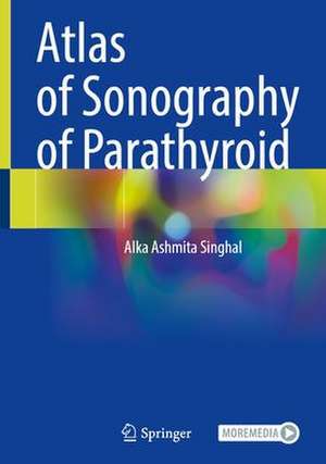
Atlas of Sonography of Parathyroid
Autor Alka Ashmita Singhalen Limba Engleză Hardback – iul 2023
Chapters cover tips and tricks in scanning along with various pitfalls. Additionally, it covers parathyroid nodules diagnosed on ultrasound in Sestamibi negative cases, localization of additional parathyroid nodules and parathyroid carcinoma. It covers correlation with surgical findings and includes histopathology images where relevant. The book serves as a teaching tool for radiologists, ENT surgeons, head and neck surgeons, general surgeons, endocrinologists, nephrologists, gastroenterologists, medical oncologists and radiation oncologists involved in the management of parathyroid disorders.
Preț: 1390.02 lei
Preț vechi: 1463.18 lei
-5% Nou
Puncte Express: 2085
Preț estimativ în valută:
266.01€ • 272.42$ • 221.28£
266.01€ • 272.42$ • 221.28£
Carte tipărită la comandă
Livrare economică 14-20 martie
Preluare comenzi: 021 569.72.76
Specificații
ISBN-13: 9789811979187
ISBN-10: 9811979189
Pagini: 183
Ilustrații: XLII, 183 p. 800 illus. in color.
Dimensiuni: 178 x 254 mm
Greutate: 0.48 kg
Ediția:2023
Editura: Springer Nature Singapore
Colecția Springer
Locul publicării:Singapore, Singapore
ISBN-10: 9811979189
Pagini: 183
Ilustrații: XLII, 183 p. 800 illus. in color.
Dimensiuni: 178 x 254 mm
Greutate: 0.48 kg
Ediția:2023
Editura: Springer Nature Singapore
Colecția Springer
Locul publicării:Singapore, Singapore
Cuprins
Introduction to parathyroid sonography.- Sonography of a typical parathyroid adenoma : Solitary parathyroids as seen on ultrasound.- Atypical Sonographic appearances of enlarged Parathyroids.- Sonography of Large parathyroid adenomas.- Role of ultrasound in evaluation of ectopic parathyroids glands.- Sonographic technique and tips to localize tiny parathyroids.- Sonography of Dual and Multiple parathyroid adenomas in sporadic Primary Hyperparathyroidism.- Role of sonography in Hyperparathyroidism in chronic kidney disease and post renal transplant recipients.-Sonography findings in Parathyroid carcinoma and Heterogenous parathyroids.- Ultrasound evaluation of Parathyroid adenoma with co-exist thyroid ds.- Ultrasound essentials in MEN Syndromes and familial Hyperparathyroidism.- Essentials of Surgical perspectives of parathyroid surgery.
Notă biografică
Dr Alka A Singhal a post graduate in radiodiagnosis from Sawai Man Singh Medical College Jaipur, affiliated to University of Rajasthan. She has worked in Sydney Australia and in Toronto Canada in the field of ultrasound Imaging. She is presently the associate director, Medanta Division of Radiology and Nuclear Medicine at Medanta the Medicity Hospital Gurgaon Delhi NCR India. Dr Singhal is a regular invited faculty for her pioneering work on the art of localizing the tiny parathyroid nodules on ultrasound and for a precise thyroid and neck lymph node ultrasound assessment.
She has more than 50 peer reviewed international publications and has delivered numerous oral and poster presentations in various international meetings related to radiology and head and neck, thyroid and parathyroid imaging. She is a member of the Indian Society of thyroid Surgeons (ISTS) and International Federation of Head and Neck oncology (IFHNO). She is a leading Thyroid, Parathyroid and Neck ultrasound imaging professional and is sought after for expert further ultrasound opinion in challenging cases. Member of IRIA, ICRI, RSNA, ECR, BISI she is a reviewer with many leading international journals and has reviewed over 400 articles. She was invited as a Scientific Chairperson in 2019 by both, European Congress of Radiology, Vienna, Austria and Radiological Society of North America, Chicago, USA, for a session on neck-thyroid and parathyroid imaging. She is a recipient of Bharat Ratna Dr Radhakrishnan Gold Medal Award 2019 for her outstanding achievements in the field of medicine and research for radiology and radiodiagnosis by the Global Economic Progress and Research Association, New Delhi.
She has more than 50 peer reviewed international publications and has delivered numerous oral and poster presentations in various international meetings related to radiology and head and neck, thyroid and parathyroid imaging. She is a member of the Indian Society of thyroid Surgeons (ISTS) and International Federation of Head and Neck oncology (IFHNO). She is a leading Thyroid, Parathyroid and Neck ultrasound imaging professional and is sought after for expert further ultrasound opinion in challenging cases. Member of IRIA, ICRI, RSNA, ECR, BISI she is a reviewer with many leading international journals and has reviewed over 400 articles. She was invited as a Scientific Chairperson in 2019 by both, European Congress of Radiology, Vienna, Austria and Radiological Society of North America, Chicago, USA, for a session on neck-thyroid and parathyroid imaging. She is a recipient of Bharat Ratna Dr Radhakrishnan Gold Medal Award 2019 for her outstanding achievements in the field of medicine and research for radiology and radiodiagnosis by the Global Economic Progress and Research Association, New Delhi.
Textul de pe ultima copertă
This comprehensive atlas covers various common and uncommon cases of parathyroid disorders encountered in the day to day radiological practice. It serves as an illustrative database with numerous state of the art images of the diverse imaging features of parathyroid nodules and their correlation with various other imaging modalities. The book includes videos as well. It covers the typical and atypical ultrasound features of parathyroid nodules, their color Doppler findings and Sestamibi correlation. Size of the nodules detected range from small sub-centimeter nodules measuring between 6-15 mm, where often the sestamibi scan is negative, and diagnostic ultrasound scan is quite vital in management of these cases. The larger nodules range in size to be even larger than the thyroid gland and completely compressing and displacing the thyroid gland, which often leads to a missed diagnosis on initial imaging.
Chapters cover tips and tricks in scanning along with various pitfalls. Additionally, it covers parathyroid nodules diagnosed on ultrasound in Sestamibi negative cases, localization of additional parathyroid nodules and parathyroid carcinoma. It covers correlation with surgical findings and includes histopathology images where relevant.
The book serves as a teaching tool for radiologists, ENT surgeons, head and neck surgeons, general surgeons, endocrinologists, nephrologists, gastroenterologists, medical oncologists and radiation oncologists involved in the management of parathyroid disorders.
Caracteristici
Focuses on imaging of typical to atypical cases of parathyroid disorders with ample images and videos Includes clinical background & presentation of the case & highlights common pitfalls of various diagnostic modalities Elucidates correlation with contrast CT and SPECT, and postoperative histopathology findings, where applicable
