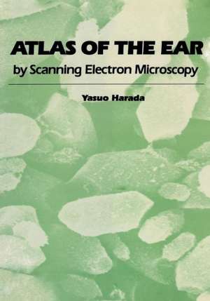
Atlas of the Ear: By Scanning Electron Microscopy
Autor Yasuo Haradaen Limba Engleză Paperback – 2 noi 2011
Preț: 716.28 lei
Preț vechi: 753.97 lei
-5% Nou
Puncte Express: 1074
Preț estimativ în valută:
137.10€ • 148.98$ • 115.24£
137.10€ • 148.98$ • 115.24£
Carte tipărită la comandă
Livrare economică 22 aprilie-06 mai
Preluare comenzi: 021 569.72.76
Specificații
ISBN-13: 9789400966000
ISBN-10: 9400966008
Pagini: 244
Ilustrații: 231 p.
Dimensiuni: 178 x 254 x 17 mm
Greutate: 0.43 kg
Ediția:Softcover reprint of the original 1st ed. 1983
Editura: SPRINGER NETHERLANDS
Colecția Springer
Locul publicării:Dordrecht, Netherlands
ISBN-10: 9400966008
Pagini: 244
Ilustrații: 231 p.
Dimensiuni: 178 x 254 x 17 mm
Greutate: 0.43 kg
Ediția:Softcover reprint of the original 1st ed. 1983
Editura: SPRINGER NETHERLANDS
Colecția Springer
Locul publicării:Dordrecht, Netherlands
Public țintă
ResearchCuprins
The Middle Ear.- The tympanic membrane.- The auditory ossicles.- The auditory ossicular chain.- The auditory muscles.- The mucous membrane of the middle ear.- The round window membrane.- The Eustachian tube.- The Inner Ear and Vestibular Organs.- The inner ear.- The otolithic organs.- The function of the otolithic organs.- The shape and composition of otoconia.- The otolithic membrane.- The formation area of the otoconia.- The absorption area of the otoconia.- The sensory cells.- The striola.- The sensory area of the macula.- The sensory cell population of the macula.- The semicircular canals.- The function of the semicircular canals.- The polarity of the vestibular organs.- The cupula.- The sensory epithelium of the crista.- The shape of the cristae ampullares.- Animals without eminentia cruciata.- The crista neglecta.- The planum semilunatum.- The transitional epithelium.- Vestibular supporting cells.- Vestibular dark cells.- Vestibular wall cells.- The calibre and number of the vestibular nerve fibres.- The cochlear aqueduct.- The endolymphatic sac.- The Cochlea.- The organ of Corti.- The outer and the inner hairs.- The tectorial membrane.- Reissner’s membrane.- The stria vascularis.- The mesothelial cell.- The spiral ganglion.- The innervation of the organ of Corti.- The nerve endings.- The vascular system of the inner ear.- Morphological Changes of the Middle and Inner Ear.- Morphological changes of the vestibular organ by aminoglycosides.- Morphological changes of the middle ear mucosa in sensory otitis media.- Cholesteatoma.- Morphological changes of the organ of Corti by aminoglycosides.- Acoustic damage to the organ of Corti.- Pathological changes of human vestibular organs.


