Cantitate/Preț
Produs

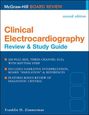
Clinical Electrocardiography: Review & Study Guide, Second Edition

Autor Franklin Zimmerman
en Limba Engleză Paperback – 16 apr 2004
Publisher's Note: Products purchased from Third Party sellers are not guaranteed by the publisher for quality, authenticity, or access to any online entitlements included with the product.


The most effective self-assessment tool for any clinician who interprets ECGs!
This unique resource offers 200 full-sized, three-channel ECGs with lead II rhythm strips, in a format that parallels the cardiology board exam. Each ECG is accompanied by a brief clinical history and followed by a narrative interpretation and board simulation, along with references for further study. A special bonus is the comprehensive section that provides diagnostic criteria for common electrocardiographic diagnoses.
Features: 
*Perfect for boards in cardiology, critical care, and anesthesiology–or for clinical practice
*200 full-size, three-channel ECGs with rhythm strip
*Each ECG accompanied by a brief clinical history in board format
*Narrative and board-type interpretations on facing page
*ECGs range from simple to complex, reflecting conditions both common and rare
*Review of diagnostic criteria for common electrocardiographic diagnoses included
*References provided for further research or study
The ultimate study aid for certification, re-certification, CME–or as a clinical refresher–this unique skill- and knowledge-building tool will help you to hone your skills in interpreting cardiac arrhythmias and other electrocardiographic abnormalities, as well as help you to effectively correlate ECG data with clinical information. More than just a study guide, this one-of-a-kind resource includes reference material that every electrocardiographer will find useful in daily clinical practice.
Citește tot Restrânge

Preț: 72331 lei

Preț vechi: 76139 lei
-5% Nou

Puncte Express: 1085

Preț estimativ în valută:
13844 14429$ 11525£

Carte tipărită la comandă

Livrare economică 06-20 ianuarie 25

Preluare comenzi: 021 569.72.76

Specificații

ISBN-13: 9780071423021
ISBN-10: 0071423028
Pagini: 421
Dimensiuni: 213 x 274 x 17 mm
Greutate: 1 kg
Ediția:2
Editura: McGraw Hill Education
Colecția McGraw Hill / Medical
Locul publicării:United States
Cuprins

Table of Electrocardiographic Diagnoses

I. RHYTHM ABNORMALITIES

A.Supraventricular Rhythms and Complexes

1.Sinus rhythm

2.Sinus arrhythmia

3.Sinus bradycardia

4.Sinus tachycardia

5.Wandering atrial pacemaker within the sinus node

6.Wandering atrial pacemaker to the AV junction

7.Sinus arrest or pause

8.Sinoatrial exit block

9.Ectopic atrial rhythm

10.Atrial premature complexes, normally conducted

11.Atrial premature complexes, aberrantly conducted

12.Atrial premature complexes, nonconducted

13.Multifocal atrial rhythm

14.Multifocal atrial tachycardia

15.Atrial tachycardia, regular 1:1 conduction, sustained

16.Atrial tachycardia, regular 1:1 conduction, short paroxysms

17.Atrial tachycardia, with non-1:1 conduction (with block)

18.Supraventricular tachycardia, unspecified

19.Atrial flutter

20.Atrial fibrillation

B.AV Junctional Rhythms and Complexes

21.AV junctional rhythm

22.AV junctional escape rhythm

23.AV junctional rhythm, accelerated

24.AV junctional escape complexes

25.AV junctional premature complexes

C.Ventricular Rhythms and Complexes

26.Ventricular premature complex(es), uniform

27.Ventricular premature complex(es), multiform

28.Ventricular premature complexes, paired

29.Ventricular parasystole

30.Ventricular tachycardia

31.Accelerated idioventricular rhythm

32.Ventricular fibrillation

33.Torsades de pointes

D.Pacemaker Function, Rhythms, and Complexes

34.Single-chamber atrial pacing

35.Single-chamber pacemaker, ventricular pacing on demand

36.Single-chamber pacemaker, ventricular pacing with complete control

37.Dual-chamber pacemaker, atrial sensing with ventricular pacing

38.Dual-chamber pacemaker, atrial and ventricular sensing and pacing

39.Pacemaker malfunction, failure to capture atrium or ventricle appropriately

40.Pacemaker malfunction, failure to sense atrial or ventricular complexes appropriately

41.Pacemaker malfunction, failure to fire appropriately on demand (inappropriate sensing of stimuli or complex)

II.AV CONDUCTION ABNORMALITIES

42.AV block, first-degree

43.AV block, second-degree, Mobitz I (Wenckebach)

44.AV block, second-degree, Mobitz II

45.AV block, second-degree, 2:1

46.AV block, high-grade

47.AV block, third-degree or complete

48.Accelerated AV conduction (short PR interval

pattern with normal QRS duration in sinus rhythm)

49.Ventricular preexcitation (WPW pattern)

50.Physiologic AV conduction delay associated with supaventricular tachyarrhythmias.

51.Nonphysiologic AV conduction delay associated with su praventricular tachyarrhythmias

III.MISCELLANEOUS AV RELATIONSHIPS

52.Ventriculophasic sinus arrhythmia

53.AV dissociation

54.Reciprocal (echo) complexes

55.Retrograde atrial activation from a ventricular focus

56.Fusion complexes

57.Ventricular capture complexes

58.Interpolation of ventricular premature complexes

IV.P-WAVE ABNORMALITIES

59.Right atrial abnormality

60.Left atrial abnormality

61.Biatrial abnormality

62.Nonspecific atrial abnormality

63.PR depression

V.ABNORMALITIES OF QRS AXIS OR VOLTAGE

64.Left axis deviation

65.Right axis deviation

66.Poor R-wave progression

67.Low voltage, limb leads

68.Low voltage, precordial leads

69.Electrical alternans

VI.INTRAVENTRICULAR CONDUCTION ABNORMALITIES,

70.Right bundle branch block, complete

71.Right bundle branch block, incomplete

72.Left anterior fascicular block

73.Left posterior fascicular block

74.Left bundle branch block, complete

75.Left bundle branch block, incomplete

76.Intraventricular conduction delay, nonspecific (includes IVCD associated with chamber enlargement)

77.Probable aberrant intraventricularconduction associated with supraventricular arrhythmia

VII.VENTRICULAR HYPERTROPHY OR ENLARGEMENT

78.Left ventricular hypertrophy by voltage criteria, with or without associated ST-T-wave abnormalities

79.Right ventricular hypertrophy

80.Combined ventricular hypertrophy

VIII.Q-WAVE MYOCARDIAL INFARCTION

81.Anteroseptal, acute or recent

82.Anteroseptal, old or of indeterminate age

83.Anterior, acute or recent

84.Anterior, old or of indeterminate age

85.Anterolateral, acute or recent

86.Anterolateral, old or of indeterminate age

87.Extensive anterior, acute or recent

88.Extensive anterior, old or of indeterminate age

89.Lateral or high lateral, acute or recent

90.Lateral or high lateral, old or of indeterminate age

91.Inferior or diaphragmatic, acute or recent

92. Inferior or diaphragmatic, old or of indeterminate age

93. Posterior, acute or recent,

94.Posterior, old or of indeterminate age

95.Suggestive of ventricular aneurysm

IX.ST-, T-, U-WAVE ABNORMALITIES

96.Normal variant, isolated J-point elevation (early repolarization pattern)

97.Isolated J-point depression

98.Normal variant, RSR’ pattern lead V1

99.Normal variant, persistent juvenile T-wave pattern

<3>100.ST- and/or T-wave abnormalities suggesting acute or recent myocardial injury

101.ST- and/or T-wave abnormalities suggesting either reciprocal change or myocardial ischemia in the setting of acute myocardial injury

102.ST- and/or T-wave abnormalities suggesting myocardial ischemia in the absence of acute myocardial injury

103.ST- and/or T-wave abnormalities associated with ventricular hypertrophy

104.ST- and/or T-wave abnormalities associated with ventricular conduction abnormality

105.ST- and/or T-wave abnormalities suggesting early, acute pericarditis

106.Nonspecific ST- and/or T-wave abnormalities

107.Post extrasystolic T-wave abnormality

108.Peaked T waves

109.Prolonged QT interval for heart rate (QTc)

110.Prominent U waves

111.Inverted U waves

X.TECHNICAL PROBLEMS

112.Incorrect electrode placement

113.Artifact secondary to tremor