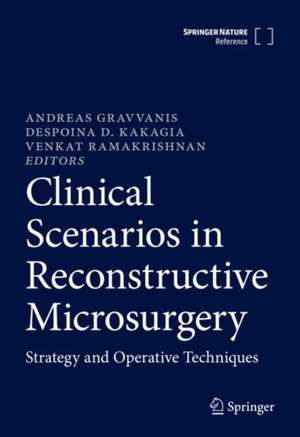
Clinical Scenarios in Reconstructive Microsurgery: Strategy and Operative Techniques
Editat de Andreas Gravvanis, Despoina D. Kakagia, Venkat Ramakrishnanen Limba Engleză Hardback – 27 noi 2022
This book presents100 cases covering all areas of reconstructive surgery. Divided into six parts (Head/Neck, Upper Extremity, Lower Extremity, Trunk, Breast and Lymphedema), it guides the reader through the difficult path of problem diagnosis, analysis and decision-making, presenting concrete steps and techniques for the successful management of patients with specific reconstructive needs. Each full-color case starts with a patient profile and continues with the diagnosis, key decisions, treatment plan, surgical procedure(s) and technical steps, postoperative management, outcome and case conclusion. Further, each case includes a discussion of pros and cons, comments, learning points and suggestions for further reading.
This book will be useful for all surgeons actively involved or interested in Reconstructive Microsurgery and valuable for Senior Residents and Fellows in Plastic, Head and Neck, Breast Surgery and Orthopedics.
Preț: 6414.38 lei
Preț vechi: 6751.97 lei
-5% Nou
Puncte Express: 9622
Preț estimativ în valută:
1227.78€ • 1334.09$ • 1032.00£
1227.78€ • 1334.09$ • 1032.00£
Carte disponibilă
Livrare economică 31 martie-14 aprilie
Preluare comenzi: 021 569.72.76
Specificații
ISBN-13: 9783030237059
ISBN-10: 3030237052
Pagini: 1000
Ilustrații: XXXIII, 1083 p. 936 illus., 869 illus. in color.
Dimensiuni: 178 x 254 mm
Greutate: 2.5 kg
Ediția:1st ed. 2022
Editura: Springer International Publishing
Colecția Springer
Locul publicării:Cham, Switzerland
ISBN-10: 3030237052
Pagini: 1000
Ilustrații: XXXIII, 1083 p. 936 illus., 869 illus. in color.
Dimensiuni: 178 x 254 mm
Greutate: 2.5 kg
Ediția:1st ed. 2022
Editura: Springer International Publishing
Colecția Springer
Locul publicării:Cham, Switzerland
Cuprins
I. HEAD AND NECK
A. SCALP RECONSTRUCTION
1. Total Scalp Replantation.
2. Scalp cutaneous defects with bone exposure following scald burn; Reconstruction with free ALT flap.
3. Scalp and calvarial bone defect following oncological resection; Reconstruction with hydroxyapatite bone grafts and free radial forearm flap.
4. Scalp and calvarial bone and dural defect following oncological resection; Reconstruction with fascia lata graft, hydroxyapatite bone graft and free latissimus dorsi flap.
5. Calvarial bone radionecrosis/osteomyelitis management; Radical debridement and free rectus muscle flap. 6. Cranial base reconstruction.
B. NASAL RECONSTRUCTION
7. Nasal skin defects resurfacing with a free flap
8. Total nasal reconstruction with radial forearm flap and a rib graft.
9. Total nasal reconstruction with radial forearm flap, forehead flap and a rib graft. 10. Nasal septum reconstruction following oncological resection.
11. Microsurgical Reconstruction of the Nasal Ala using a Composite Auricular Graft Based on the Superficial Temporal Vessels.
C. TONGUE RECONSTRUCTION
12. Partial glossectomy reconstruction with ulnar forearm flap.
13. Subtotal glossectomy reconstruction with saphenous flap.
14. Tongue reconstruction with ALT/vastus lateralis after total glossectomy glossectomy.
D. LIP RECONSTRUCTION
15. Total lower lip reconstruction with a skin flap and a tendon graft.
16. Total lower lip reconstruction with functioning free muscle.
E. EAR RECONSTRUCTION
17. Ear replantation.
18. Ear reconstruction using microsurgical techniques.
F. MID FACE RECONSTRUCTION
19. Massive facial defect following trauma.
20. Mid face reconstruction with TAP flap. 21. Maxillary and mid- face reconstruction with scapular flap.
22. Mandibular reconstruction with CAD/CAM technology.
23. Mandible osteomyletis reconstruction.
24. Reconstruction of Temporomandibular Joint with a Fibula Free Flap: A Case Report With a Histological Study.
25. Mandibular reconstruction with iliac crest. 26. Immediate oromandibular defect reconstruction with double flap (fibula and ALT).
27. Immediate oromandibular defect reconstruction with chimeric fibula flap.
28. Head and Neck secondary reconstruction in a vessel-depleted neck.
29. Craniofacial deformity reconstruction.
30. Hemifacial atrophy reconstruction with parascapular flap.
31. Noma disease reconstruction.
32. Facial transplantation.
G. UPPER DIGESTIVE SYSTEM RECONSTRUCTION
33. Hypopharynx reconstruction.
34. Robotic flap inset in pharyngeal reconstruction.
35. Oesophageal reconstruction with free cutaneous flap.
36. Oesophageal reconstruction with supercharged ileocolon flap.
37. Repair of a large pharyngocutaneous fistula with a free jejunal patch flap after salvage laryngectomy. 38. Voice reconstruction with free ileocolon flap transfer.
39. Tracheal reconstruction.
40. Tracheal transplantation.
H. FACIAL PARALYSIS
41. Adult Facial nerve palsy reconstruction. Gracilis functioning muscle innervated with cross face nerve graft.
42. Microsurgical eye reanimation.
43. One-stage reconstruction of facial paralysis using latissimus dorsi functioning muscle. 44. One-stage reconstruction of facial paralysis using masseter nerve-innervated gracilis.
45. One-stage reconstruction of facial paralysis using spinal accessory nerve-innervated gracilis.
46. Adult Facial nerve palsy reconstruction. Double innervation of Gracilis functioning muscle innervated with cross face nerve graft and masseter nerve.
47. Synkinesis treatment following nerve palsy.
48. Congenital facial nerve palsy (Moebius syndrome).
II. UPPER EXTREMITY
A. BRACHIAL PLEXUS INJURIES - PERIPHERAL NERVE INJURIES49. Obstetric brachial plexus injuries
50. Adult brachial plexus immediate reconstruction
With intercostal nerves.
51. Adult brachial plexus immediate reconstruction
With contralateral C7.
52. Infraclavicular brachial plexus injury.
53. Brachial plexus secondary reconstruction with functional muscle (elbow flexion-finger extension).
54. Brachial plexus secondary reconstruction with functional muscle (shoulder abduction).
55. Spaghetti wrist injury.
56. Ischemic compartment functional reconstruction.
57. Secondary reconstruction of high median nerve injury Motor and Sensory Nerve Transfers to Restore Function.
58. Application of free temporoparietal fascial flap for recurrent neural adhesion of superficial radial nerve.
59. Painful neuroma reconstruction.
B. THUMB - HAND RECONSTRUCTION
60. Thumb amputation and replantation. 61. Multiple finger replantations.
59. Thumb reconstruction.
60. Thumb pulp reconstruction with sensate flap.
61. Thumb pulp reconstruction with great toe hemi-pulp flap transfer.
62. Thumb reconstruction with toe to thumb.
63. Thumb reconstruction with wrap around flap.
64. Congenital thumb reconstruction with index pollicisation.
65. Thumb and the first web reconstruction with neurovascularised instep flap.
66. Thumb reconstruction with medial plantar artery perforator flaps.
67. Thumb reconstruction with medial femoral corticoperiosteal free flap.
68. The free instep flap for palmar and digital resurfacing.
69. Degloving injuries.
70. Transverse sensate thoracodorsal artery perforator flaps to resurface fingers.
71. Free distal forearm perforator flap for digital reconstruction.
72. SUBPRA for volar digital resurfacing.
73. Dorsal digital sensate free flap for reconstruction of volar soft tissue defect of digits.
74. Free temporoparietal flap in hand coverage.
75. Free serratus anterior fascia flap for reconstruction of hand and finger defects.
76. Thenar reconstruction with functional gracilis flap.
77. Hand skin defects with tendon defect.
78. Volar defects in fingers with distal ulnar artery perforator flaps.
79. Bilateral hand transplant.
III. TRUNK
80. Large chest wall defects.
81. Post burn scar contracture.
82. Axillary reconstruction with ALT.
83. Functional reconstruction of abdominal wall defect.
84. Pelvic floor reconstruction with Latissimus dorsi.
85. Vaginal reconstruction.
86. Penile reconstruction.
87. Functional bladder reconstruction.
88. Breast and chest wall reconstruction with the transverse musculocutaneous gracilis flap in Poland syndrome.
89. Autologous reconstruction of a complex form of Poland syndrome using 2 abdominal perforator free flaps.
90. Aesthetic restoration of Poland's syndrome in a male patient using free anterolateral thigh perforator flap as autologous filler.
IV. BREAST
91. Areola sparing mastectomy and immediate DIEP flap.
92. Skin sparing mastectomy and TUG flap.
93. Secondary breast reconstruction with SGAP flap P.
94. Tertiary reconstruction (previous failed implant reconstruction) with PAP flap.
95. Case of a failed free DIEP, rexploration and outcome.
96. Stacked abdominal flap for unilateral breast reconstruction.
97. Breast reconstruction with Lumbar artery perforator flap.
98. Breast reconstruction with SIEA flap.
99. Breast reconstruction with ALT.
100. Breast reconstruction with Vertical Posteromedial thigh flap.
101. Four free flaps for 'stacked' bilateral breast reconstruction.
102. Immediate breast reconstruction with a free flap and an implant. 103. Partial mastectomy reconstruction with TDAP flap.
V. LYMPHEDEMA
104. Lymphatico-venous anastomoses for lower extremity stage I lymphedema. 105. Vascularised lymph node transfer for upper extremity stage II lymphedema.
106. Secondary breast reconstruction with DIEP flap and lymph node transfer.
107. Fibro-Lipo-Lymph-Aspiration With a Lymph Vessel Sparing Procedure to Treat Advanced Lymphedema After Multiple Lymphatic-Venous Anastomoses.
108. Vascularized lymph node transfer from thoracodorsal axis for congenital lymphedema of the lower limb.
109. Lymphatic vessel grafting for prevention of venous reflux into a sclerotic lymphatic vessel in supermicrosurgical lymphaticovenular anastomosis.
VI. LOWER EXTREMITY
110. Free fibular transfer for femoral head osteonecrosis. 111. Femoral bone reconstruction following oncological resection.
112. Coverage of knee joint/ arthroplasty prosthesis exposure with free flap.
113. Coverage of knee joint with supercharged pedicled perforator flap.
114. Gustillo type IIIB tibial defects.
115. Gustillo type IIIC tibial defects; reconstruction with a flow-through free flap.
116. Peroneal nerve palsy - functional reconstruction.
117. Tibial bone defect reconstruction with Illizarov and free flap.
118. Tibial bone defects reconstruction with free fibula flaps.
119. Heel defect reconstruction.
120. Achilles tendon reconstruction.
121. Plantar weight bearing area defects.
122. Supermicrosurgical free sensate superficial circumflex iliac artery perforator flap for reconstruction of a soft tissue defect of the ankle.
123. Reconstruction of metatarsal bone defects with a free fibular flap.
124. Lower limb transplantation.
125. Reconstruction of the ankle complex wound with a fabricated superficial circumflex iliac artery chimeric flap including the sartorius muscle.
126. Management of sciatic nerve schwannoma.
Notă biografică
Andreas Gravvanis currently serves as Head of Plastic, Reconstructive and Aesthetic Surgery in the Metropolitan Hospital of Athens. Following his basic plastic surgery training at the General State Hospital of Athens and at Chang Gung Memorial Hospital, Taiwan, where he focused on facial paralysis/head and neck reconstruction (sponsored by MILLESI Award), he accomplished three microsurgical fellowships at St Andrews Centre for Plastic Surgery, UK, focusing on breast/microsurgery; Gent Plastic Surgery University Department, Belgium, focusing on breast reconstruction (sponsored by EURAPS Scholarship); and EURAPS/AAPS fellowship at MD Anderson Cancer Centre focusing on head and neck cancer reconstruction. Gravvanis was appointed as a Consultant Plastic Surgeon with special interest in Breast Reconstruction in Queen Victoria Hospital, East Grinstead, UK, for the period 2007–2008. He was subsequently appointed as a Consultant in Plastic Surgery at General State Hospital of Athens, 2009–2017. His interests include all aspects of microsurgery dealing with cancer and trauma reconstruction along with facial palsy. He also focuses on facial and breast aesthetic surgery. Three-dimensional (3D) photography and 3D virtual models are an integral part of his practice in both reconstructive and aesthetic surgery. Gravvanis is the author and co-author of 62 publications in peer-reviewed journals with over a 1000 citations and 3 book chapters. He is the editor of the reference book Clinical Scenarios in Reconstructive Microsurgery published by Springer. He is also an active member of EURAPS, a member of the editorial board of Microsurgery, and a reviewer for Aesthetic Plastic Surgery.
Despoina D. Kakagia is Professor of Plastic Surgery at Democritus University in Thrace, Greece, and has been practicing reconstructive and cosmetic plastic surgery since 1998. Following her comprehensive training in plastic surgery at Metaxa Cancer Institute and Athens General State Hospital in Greece, she underwent further training in microsurgery of the hand and breast at Canniesburn, Glasgow, Scotland; Chelsea and Westminster Hospital, London, UK; and Aachen University Hospital, Germany.
Professor Kakagia is a member of the Hellenic Society of Plastic Reconstructive and Aesthetic Surgery (HESPRAS), the Hellenic Society of Reconstructive Microsurgery and Hand and Upper Limb Surgery, and the Hellenic Society of Wound Healing and is an active member of EURAPS. Her special interests include oncologic surgery and trauma, breast surgery, burns, and wound healing.
Professor Kakagia has been teaching plastic surgery at undergraduate and postgraduate level since 2006 and has participated as an instructor in many microsurgery training courses. She is the author or co-author of more than 70 publications in peer-reviewed international journals and has authored 1 book as well as 15 book chapters. She is an active reviewer in more than 30 international medical journals.
Venkat Ramakrishnan is a Consultant Plastic Surgeon at the St Andrew’s Centre for Plastic Surgery and Burns, Chelmsford, United Kingdom, and Visiting Professor of Microsurgery at the Anglia Ruskin University, Chelmsford, United Kingdom. His main area of work is microsurgical reconstruction of the breast and aesthetic surgery of the breast.
Ramakrishnan plays a major role as a trainer in microsurgery and was the inaugural Tutor in Plastic Surgery at the Royal College of Surgeons of England, London. He was the Director of the St Andrew’s Centre and has had roles in the BAPRAS Council and the project board of the National Mastectomy and Reconstruction audit. He is a member of the editorial board of the Journal of Plastic, Reconstructive and Aesthetic Surgery and Archives of Plastic Surgery. He is a fellow of the Royal College of Surgeons of England and the Royal Australasian College of Surgeons.
Ramakrishnan has numerous publications and presentations at national and international meetings. His main areas of research and audit are in microsurgical techniques, service delivery, and microcirculation in free flaps.
Caracteristici
Focuses on very specific and often complex reconstructive problems
Thoroughly analyzes the decision-making steps for each case
Features accompanying high resolution photos for each case
Thoroughly analyzes the decision-making steps for each case
Features accompanying high resolution photos for each case
