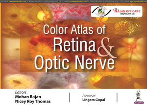
Color Atlas of Retina & Optic Nerve
Autor Mohan Rajan, Nicey Roy Thomasen Limba Engleză Hardback – apr 2022
Comprising more than 500 archetypal images, each disease is clearly illustrated showing clinical features and signs, from its early to later stages.
The images are followed by a brief description highlighting key characteristics of the disease, providing clinicians with a comprehensive overview and enabling them to diagnose ocular conditions with ease.
Divided into 15 sections, the book begins with an introduction to the ‘normal’ fundus. The following sections examine different retinal disorders, from retinal degeneration, uveitis, and infections, to traumatic chorioretinopathy and optic disc anomalies.
The atlas concludes with chapters on ocular oncology and complications of surgery.
A complete section is dedicated to paediatric retinal diseases.
Preț: 609.50 lei
Preț vechi: 641.58 lei
-5% Nou
116.64€ • 126.66$ • 97.98£
Carte disponibilă
Livrare economică 02-16 aprilie
Livrare express 18-22 martie pentru 78.51 lei
Specificații
ISBN-10: 9354651135
Pagini: 370
Dimensiuni: 286 x 222 x 25 mm
Greutate: 1.74 kg
Editura: Jp Medical Ltd
Colecția Jaypee Brothers Medical Publishers
Locul publicării:Delhi, India
Cuprins
- NORMAL FUNDUS
- RETINAL DEGENERATIONS AND DYSTROPHIES
- PAEDIATRIC RETINAL DISEASES
- RETINAL VASCULAR DISEASE
- CHOROIDAL VASCULAR/BRUCH'S MEMBRANE DISEASE
- CENTRAL SEROUS CHORIORETINOPATHY
- INFLAMMATORY DISEASE/UVEITIS
- INFECTIONS
- EPIRETINAL MEMBRANE, VITREOMACULAR TRACTION, MACULAR HOLE
- VITREOUS DEGENERATION
- TRAUMATIC CHORIORETINOPATHY
- PERIPHERAL RETINAL DEGENERATIONS AND RHEGMATOGENEOUS RETINAL DETACHMENT
- OPTIC DISC ANOMALIES AND DISEASES
- ONCOLOGY
- COMPLICATIONS OF OCULAR SURGERY
Notă biografică
Mohan Rajan MBBS DO Dip NB MNAMS FMRF MCh FACS FIAMS FRCS DSc PhD
Nicey Roy Thomas MBBS MS FICO FMRF
Descriere
This atlas is a practical guide to the diagnosis of common retina and optic nerve disorders.
Comprising more than 500 archetypal images, each disease is clearly illustrated showing clinical features and signs, from its early to later stages.
The images are followed by a brief description highlighting key characteristics of the disease, providing clinicians with a comprehensive overview and enabling them to diagnose ocular conditions with ease.
Divided into 15 sections, the book begins with an introduction to the ‘normal’ fundus. The following sections examine different retinal disorders, from retinal degeneration, uveitis, and infections, to traumatic chorioretinopathy and optic disc anomalies.
The atlas concludes with chapters on ocular oncology and complications of surgery.
A complete section is dedicated to paediatric retinal diseases.
