Colorectal Tumors: Atlas of Large Section Histopathology
Autor Tibor Toten Limba Engleză Hardback – 14 iun 2005
Large-section histopathology widens your perspectives...
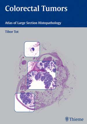
Correct diagnosis and staging are essential in determining the appropriate therapy of colorectal carcinoma, one of the most common malignancies in America and Europe. As medical science continues to develop rapidly, histopathology remains an essential part of diagnosis in most malignant diseases. In this era of interdisciplinary medicine, the role of pathology has expanded to provide images that easily correlate with endoscopic, radiological, or operative findings.
In this remarkable atlas, Tibor Tot presents colon pathology in large histological sections, with cross-sections of entire tumors in their anatomic environments and their circumferential surgical margins. These unique images form a guide to diagnosis, tumor typing and staging according to TNM criteria. They help to assess the completeness of a surgical excision and to understand the heterogeneity of colorectal carcinomas.
Features:
- Cases illustrated in two-page spreads with clinical information, conventional histopathology, and large-section histology images enlarged to almost a full page.
- Pathology seen in the context of surrounding tissues
- The margins of malignant tumors visible in their entirety
- Schematic guides to interpretation of the large-section images
- Emphasis on diagnostic advantages of using large section technique
- Technical guidelines for obtaining large-section histopathology specimens
Preț: 841.38 lei
Preț vechi: 903.65 lei
-7% Nou
Puncte Express: 1262
Preț estimativ în valută:
161.02€ • 167.48$ • 132.93£
161.02€ • 167.48$ • 132.93£
Carte tipărită la comandă
Livrare economică 11-23 aprilie
Preluare comenzi: 021 569.72.76
Specificații
ISBN-13: 9783131405913
ISBN-10: 3131405910
Pagini: 152
Ilustrații: 293
Greutate: 0.85 kg
Ediția:1st edition
Editura: Thieme
Colecția Thieme
ISBN-10: 3131405910
Pagini: 152
Ilustrații: 293
Greutate: 0.85 kg
Ediția:1st edition
Editura: Thieme
Colecția Thieme
Recenzii
Superbly illustrated atlas...composed of remarkable, full page color images...I am not aware of any other atlas that so beautifully illustrates this method. ...Photographs in this atlas are excellent to visualize the anatomy. ..A useful atlas to have any department playing host to junior trainees who are still building confidence with dissection.--Histopathology This is a beautifully produced atlas...The illustrations are of exceptional quality and of particular value in understanding tumor spread... This novel publication focuses on large section histopathology making pathology much more accessible, and would be of value to radiologists, oncologists, and surgeons in particular. The book would be an excellent adjunct for the teaching of colorectal pathology in a wide rande of clinical settings, including to undergraduates. [The book] is highly recommended for any radiologist with an interest in colorectal cancer and the correlation between imaging and pathology. The quality of the images justifies the expense [of the book]. It would be a worthy addition to any medical imaging departmental library.--RAD MagazineUsing large histologic sections, including representative transections of entire tumors in their anatomic environments, all the physicians involved in diagnosis or treatment can better type tumors and stage according to TNM criteria. --SciTech Book NewsAn excellent reference...a departure from the typical picture atlas[providing] a brief review of the topic using case reports and whole mount slide images to illustrate relevant pathology...useful...an excellent educational resource.--Doody's Book Reviews
Textul de pe ultima copertă
Large-section histopathology widens your perspectives...
Correct diagnosis and staging are essential in determining the appropriate therapy of colorectal carcinoma, one of the most common malignancies in America and Europe. As medical science continues to develop rapidly, histopathology remains an essential part of diagnosis in most malignant diseases. In this era of interdisciplinary medicine, the role of pathology has expanded to provide images that easily correlate with endoscopic, radiological, or operative findings.
In this remarkable atlas, Tibor Tot presents colon pathology in large histological sections, with cross-sections of entire tumors in their anatomic environments and their circumferential surgical margins. These unique images form a guide to diagnosis, tumor typing and staging according to TNM criteria. They help to assess the completeness of a surgical excision and to understand the heterogeneity of colorectal carcinomas.
Features:
Correct diagnosis and staging are essential in determining the appropriate therapy of colorectal carcinoma, one of the most common malignancies in America and Europe. As medical science continues to develop rapidly, histopathology remains an essential part of diagnosis in most malignant diseases. In this era of interdisciplinary medicine, the role of pathology has expanded to provide images that easily correlate with endoscopic, radiological, or operative findings.
In this remarkable atlas, Tibor Tot presents colon pathology in large histological sections, with cross-sections of entire tumors in their anatomic environments and their circumferential surgical margins. These unique images form a guide to diagnosis, tumor typing and staging according to TNM criteria. They help to assess the completeness of a surgical excision and to understand the heterogeneity of colorectal carcinomas.
Features:
- Cases illustrated in two-page spreads with clinical information, conventional histopathology, and large-section histology images enlarged to almost a full page.
- Pathology seen in the context of surrounding tissues
- The margins of malignant tumors visible in their entirety
- Schematic guides to interpretation of the large-section images
- Emphasis on diagnostic advantages of using large section technique
- Technical guidelines for obtaining large-section histopathology specimens


