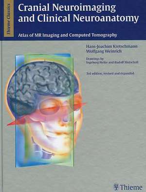
Cranial Neuroimaging and Clinical Neuroanatomy: Magnetic Resonance Imaging andComputed Tomography
Autor Hans-Joachim Kretschmann, Wolfgang Weinrichen Limba Engleză Hardback – 9 dec 2003
Written by experts in the field, this beautifully illustrated text/atlas provides the tools you need to directly visualize and interpret cranial CT and MR images. It reviews with exacting detail the normal anatomic brain structures identified on sagittal, coronal, and axial imaging planes. Use this book to make accurate and complete neurological assessments at the earliest possible stages - before reaching the sectioning or operating table.
This revised and expanded third edition contains nearly 600 illustrations - most in color - that provide graphic representations of brain structures, arteries, arterial territories, veins, nerves and neurofunctional systems. The illustrations depict anatomic structures in shades of gray similar to the way they are seen in CT and MR images.
Highlights of the third edition:
- Content and illustrations expanded by more than 20%
- High resolution T1 and T2 weighted MR images
- Improved anatomic terminology for more accurate descriptions of findings
Preț: 2417.99 lei
Preț vechi: 2545.25 lei
-5% Nou
Puncte Express: 3627
Preț estimativ în valută:
462.66€ • 483.11$ • 382.06£
462.66€ • 483.11$ • 382.06£
Carte indisponibilă temporar
Doresc să fiu notificat când acest titlu va fi disponibil:
Se trimite...
Preluare comenzi: 021 569.72.76
Specificații
ISBN-13: 9781588901453
ISBN-10: 1588901459
Pagini: 451
Ilustrații: 664
Dimensiuni: 234 x 297 x 30 mm
Greutate: 2.09 kg
Ediția:3rd edition, revised and expanded
Editura: Thieme
Colecția Thieme
ISBN-10: 1588901459
Pagini: 451
Ilustrații: 664
Dimensiuni: 234 x 297 x 30 mm
Greutate: 2.09 kg
Ediția:3rd edition, revised and expanded
Editura: Thieme
Colecția Thieme
Recenzii
'A richly illustrated state-of-the-art work in neuroanatomy...a practical and instructive diagnostic tool.' -- Surgical and Radiologic AnatomyPraise of earlier editions:'. . .the authors are to be commended for providing such a large amount of information in such a clearly organized and well-illustrated fashion. I highly recommend this book to all radiologists who interpret cross-sectional images of the brain.' --American Journal of Neuroradiology'This creatively-illustrated atlas and text. . .is full of useful material and would be an excellent addition to a neuroradiology library.' --Radiology'An excellent textbook of practical anatomy. . .a valuable reference for studying neuroanatomy. . .this text should be found in department libraries in these fields [of radiology, neuroradiology, neurology, and neurosurgery]. . .This beautifully drawn text is an excellent introduction of multiplanar neuroanatomy.' --American Journal of Roentenology
Notă biografică
Professor and Head, Department of Neuroanatomy, Medizinische Hochschule Hannover, Germany
Textul de pe ultima copertă
Written by experts in the field, this beautifully illustrated text/atlas provides the tools you need to directly visualize and interpret cranial CT and MR images. It reviews with exacting detail the normal anatomic brain structures identified on sagittal, coronal, and axial imaging planes. Use this book to make accurate and complete neurological assessments at the earliest possible stages - before reaching the sectioning or operating table.
This revised and expanded third edition contains nearly 600 illustrations - most in color - that provide graphic representations of brain structures, arteries, arterial territories, veins, nerves and neurofunctional systems. The illustrations depict anatomic structures in shades of gray similar to the way they are seen in CT and MR images.
Highlights of the third edition:
This revised and expanded third edition contains nearly 600 illustrations - most in color - that provide graphic representations of brain structures, arteries, arterial territories, veins, nerves and neurofunctional systems. The illustrations depict anatomic structures in shades of gray similar to the way they are seen in CT and MR images.
Highlights of the third edition:
- Content and illustrations expanded by more than 20%
- High resolution T1 and T2 weighted MR images
- Improved anatomic terminology for more accurate descriptions of findings
Descriere
This book is designed as a practical tool. The illustrations of the neurofunctional systems as they are localized in the tomographic planes are meant to orient the reader as to their localization in CT, MR and PET images. They also make it possible to extrapolate the clinical symptoms which correlate to the pathological CT and MR findings.
