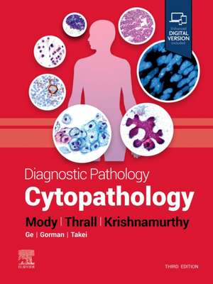
Diagnostic Pathology: Cytopathology: Diagnostic Pathology
Autor Dina R Mody, Michael J. Thrall, Savitri Krishnamurthyen Limba Engleză Hardback – 18 oct 2022
- Covers all areas of cytopathology, including clinical, radiologic, cytopathological features, immunohistochemical, and molecular correlates where applicable
- Contains new chapters on recently described entities, ancillary molecular tests specific to thyroid, prognostic/therapy-related immunomarkers in cell blocks, and small biopsies
- Provides new immunohistochemical and molecular coverage, including new immunostains and genomic targets, as well as ancillary molecular test coverage in the thyroid section
- Incorporates new reporting terminology (such as serous fluid and effusions) and updates to existing reporting terminologies
- Reflects the expanded use of small biopsies as an adjunct to or replacement for aspirations, with many more images added and updated throughout
- Keeps you up to date with current and emerging reporting systems, including pancreaticobiliary and breast
- Features more than 2,400 print and online images, including carefully annotated cytology images as well as histology and gross pathology photos, full-color illustrations, clinical photographs, and radiologic images to help practicing and in-training pathologists reach a confident diagnosis
- Includes new videos on such topics as the cytoprepratory process, cell transfer cell block and smears, collodion bag cell block, and more
- Employs consistently templated chapters, bulleted content, key facts, a variety of tables, annotated images, pertinent references, and an extensive index for quick, expert cytopathology reference at the point of care
- Includes the enhanced eBook version, which allows you to search all text, figures, and references on a variety of devices
Din seria Diagnostic Pathology
- 24%
 Preț: 1221.50 lei
 - 24%
 Preț: 1457.26 lei
 - 5%
 Preț: 1225.22 lei
 - 5%
 Preț: 1205.14 lei
 - 24%
 Preț: 1610.44 lei
 - 24%
 Preț: 1578.34 lei
 - 24%
 Preț: 1462.52 lei
 - 24%
 Preț: 1620.36 lei
 - 24%
 Preț: 1608.87 lei
 - 25%
 Preț: 1452.85 lei
 - 24%
 Preț: 1461.30 lei
 - 25%
 Preț: 1107.73 lei
 - 24%
 Preț: 1110.69 lei
 - 24%
 Preț: 1206.29 lei
 - 24%
 Preț: 1406.65 lei
 - 24%
 Preț: 1112.17 lei
 - 5%
 Preț: 1515.24 lei
 - 24%
 Preț: 1351.48 lei
 - 24%
 Preț: 1513.73 lei
 - 5%
 Preț: 1834.07 lei
 - 39%
 Preț: 1324.52 lei
 - 25%
 Preț: 1205.67 lei
 - 25%
 Preț: 1244.70 lei
 - 24%
 Preț: 1478.05 lei
 - 25%
 Preț: 1248.57 lei
Preț: 1343.10 lei
Preț vechi: 1770.46 lei
-24% Nou
257.08€ • 279.34$ • 216.09£
Carte disponibilă
Livrare economică 24 martie-07 aprilie
Livrare express 14-20 martie pentru 457.98 lei
Specificații
ISBN-10: 0323878679
Pagini: 825
Ilustrații:
Features more than 2,400 print and online images, including carefully annotated histology and gross pathology photos, full-color illustrations, clinical photographs, and radiologic images
Dimensiuni: 216 x 276 x 41 mm
Greutate: 2.5 kg
Ediția:3
Editura: Elsevier
Seria Diagnostic Pathology
Cuprins
R. Mody, MD
8 Cytopreparation, Instrumentation, and Automated
Screening in Gynecologic Cytology
Michael J. Thrall, MD and Debora A. Smith, CT(ASCP)
10 Specimen Adequacy in Cervicovaginal Cytology
Dina R. Mody, MD and George G. Birdsong, MD
SECTION 2: BENIGN AND INFECTIOUS
CONDITIONS
14 Normal Pap Test
Nikolaos Chantziantoniou, BSc, ART(CSMLS), CFIAC and Dina R. Mody, MD
16 Infectious and Other Organisms in Pap Tests
Nikolaos Chantziantoniou, BSc, ART(CSMLS), CFIAC and Dina R. Mody, MD
18 Nonneoplastic Findings, Mimics, and Artifacts
Ekene I. Okoye, MD and Dina R. Mody, MD
SECTION 3: SQUAMOUS CELL
ABNORMALITIES AND MIMICS
28 Low-Grade Squamous Intraepithelial Lesion and
Mimics
Dina R. Mody, MD
30 High-Grade Squamous Intraepithelial Lesion and
Mimics
Dina R. Mody, MD and Ekene I. Okoye, MD
34 Atypical Squamous Cells of Undetermined
Significance
Nikolaos Chantziantoniou, BSc, ART(CSMLS), CFIAC and Dina R. Mody, MD
36 Atypical Squamous Cells, Cannot Rule Out High-
Grade Squamous Intraepithelial Lesion
Dina R. Mody, MD and Nikolaos Chantziantoniou, BSc, ART(CSMLS), CFIAC
38 Squamous Cell Carcinoma of Cervix, Variants and
Mimics
Dina R. Mody, MD
SECTION 4: GLANDULAR CELL
ABNORMALITIES AND MIMICS
46 Endocervical Adenocarcinoma In Situ, Variants and
Mimics
Dina R. Mody, MD
52 Endocervical Adenocarcinoma, Variants and Mimics
Dina R. Mody, MD
56 Adenocarcinoma, Minimal Deviation
Dina R. Mody, MD
58 Endometrial Cancers: Usual Types, Variants, and Mimics
Ekene I. Okoye, MD and Dina R. Mody, MD
64 Atypical Glandular Cells: Endocervicals, Endometrials, and Glandulars, NOS
Dina R. Mody, MD
70 Endometrial Cells in Pap Test and Glandular Cells Status Post Hysterectomy
Dina R. Mody, MD and Ekene I. Okoye, MD
SECTION 5: EXTINE
CARCINOMAS AND OTHER
MALIGNANCIES OF FEMALE GENITAL
TRACT
74 Extrauterine Carcinomas and Presentations in Cervicovaginal Cytology
Dina R. Mody, MD
76 Small Cell Neuroendocrine Carcinoma of Cervix
Dina R. Mody, MD
80 Other Uncommon Malignancies in Cervicovaginal Cytology
Dina R. Mody, MD
SECTION 6: MOLECULAR TESTING IN
GYNECOLOGIC CYTOLOGY
86 HPV and Other Molecular Testing in Gynecologic Cytology
Michael J. Thrall, MD
SECTION 7: DIRECTLY SAMPLED
ENDOMETRIAL CYTOLOGY
92 Directly Sampled Endometrial Cytology
Yuko Sugiyama, MD, PhD, FIAC and Dina R. Mody, MD
SECTION 8: ANAL CYTOLOGY
98 Anal Cytology
Michael J. Thrall, MD
Part II: Exfoliative Cytopathology
SECTION 1: RESPIRATORY TRACT
INCLUDING LUNG FNAS
102 Specimen Types in Respiratory Cytology and Adequacy Criteria
Michael J. Thrall, MD
104 Benign and Reactive Changes
Michael J. Thrall, MD
108 Pneumocystis Pneumonia and Mimics
Michael J. Thrall, MD
110 Fungal Organisms in Respiratory Cytology
Michael J. Thrall, MD
112 Parasitic Organisms in Respiratory Cytology
Michael J. Thrall, MD
114 Viral Infections (Cytomegalovirus, Herpesvirus, and Others)
Michael J. Thrall, MD
116 Mycobacteria and Other Bacterial Infections
Michael J. Thrall, MD
118 Sarcoidosis and Other Immune-Related Conditions
Michael J. Thrall, MD
120 Pulmonary Alveolar Proteinosis and Mimics
Michael J. Thrall, MD
122 Miscellaneous Findings Including Contaminants
Michael J. Thrall, MD
124 Adenocarcinoma
Michael J. Thrall, MD
128 Squamous Cell Carcinoma
Michael J. Thrall, MD
130 Small Cell Carcinoma
Michael J. Thrall, MD
132 Large Cell Neuroendocrine Carcinoma
Michael J. Thrall, MD
134 Carcinoid and Atypical Carcinoid
Michael J. Thrall, MD
136 Rare Benign and Low Malignant Potential Tumors
Michael J. Thrall, MD
138 Rare Malignant Tumors
Michael J. Thrall, MD
140 Pulmonary Lymphoma
Michael J. Thrall, MD
142 Pulmonary Metastasis
Michael J. Thrall, MD
SECTION 2: GASTROINTESTINAL TRACT
146 Specimen Types in Gastrointestinal Cytology and Normal Cellular Components
Blythe K. Gorman, MD and Dina R. Mody, MD
148 Parasitic Infections
Blythe K. Gorman, MD and Dina R. Mody, MD
150 Viral Infections
Blythe K. Gorman, MD and Dina R. Mody, MD
152 Esophagitis and Barrett Esophagus
Blythe K. Gorman, MD and Dina R. Mody, MD
154 Esophageal Adenocarcinoma
Blythe K. Gorman, MD and Dina R. Mody, MD
156 Esophageal Squamous Cell Carcinoma
Blythe K. Gorman, MD
158 Gastritis and Intestinal Metaplasia
Blythe K. Gorman, MD and Dina R. Mody, MD
160 Gastric Adenocarcinoma
Blythe K. Gorman, MD and Dina R. Mody, MD
162 Gastric Lymphoma
Blythe K. Gorman, MD and Dina R. Mody, MD
164 Ampulla/Bile Duct/Pancreatic Duct Reactive
Changes
Blythe K. Gorman, MD and Dina R. Mody, MD
166 Ampulla/Bile Duct/Pancreatic Duct Adenocarcinoma
Blythe K. Gorman, MD and Dina R. Mody, MD
168 Colorectal Adenoma/Carcinoma
Blythe K. Gorman, MD and Dina R. Mody, MD
170 Neuroendocrine Tumor/Carcinoma
Blythe K. Gorman, MD and Dina R. Mody, MD
172 Spindle Cell Neoplasms of Gastrointestinal Tract,
Including Gastrointestinal Stromal Tumors
Blythe K. Gorman, MD and Dina R. Mody, MD
SECTION 3: CEREBROSPINAL FLUID
176 Normal Cerebrospinal Fluid and Contamination by Normal Elements
Hidehiro Takei, MD and Michael J. Thrall, MD
180 Infectious Meningitis
Hidehiro Takei, MD and Michael J. Thrall, MD
182 Aseptic and Mollaret Meningitis
Hidehiro Takei, MD and Michael J. Thrall, MD
184 Subarachnoid Hemorrhage
Hidehiro Takei, MD and Michael J. Thrall, MD
186 Neurodegenerative Diseases
Hidehiro Takei, MD and Michael J. Thrall, MD
188 Primary Brain Tumors
Hidehiro Takei, MD and Michael J. Thrall, MD
190 Leukemia and Lymphoma
Hidehiro Takei, MD and Michael J. Thrall, MD
192 Metastasis in CSF
Hidehiro Takei, MD and Michael J. Thrall, MD
SECTION 4: PLEURAL, PERITONEAL,
PERICARDIAL, AND PELVIC FLUID AND
WASHINGS
196 Normal Cellular Components, Reactive Mesothelial Proliferations, and Reporting Terminology
Donna M. Coffey, MD and Michael J. Thrall, MD
200 Infectious Conditions
Donna M. Coffey, MD and Michael J. Thrall, MD
202 Autoimmune Diseases
Donna M. Coffey, MD and Michael J. Thrall, MD
204 Malignant Effusion, Mesothelioma
Donna M. Coffey, MD and Michael J. Thrall, MD
208 Malignant Effusion, Carcinomas
Donna M. Coffey, MD and Michael J. Thrall, MD
212 Malignant Effusion, Sarcomas
Donna M. Coffey, MD, Nour Sneige, MD, and Michael J. Thrall, MD
214 Lymphoid Effusions and Lymphomas
Donna M. Coffey, MD and Michael J. Thrall, MD
216 Primary Effusion Lymphoma
John M. Stewart, MD, PhD and Michael J. Thrall, MD
218 Endometriosis and Endosalpingiosis
Donna M. Coffey, MD, Nour Sneige, MD, and Michael J.
Thrall, MD
220 Ovarian Neoplasms
Donna M. Coffey, MD and Michael J. Thrall, MD
222 Immunocytochemistry, Histochemistry, and Other Ancillary Techniques
Donna M. Coffey, MD and Michael J. Thrall, MD
SECTION 5: URINARY CYTOLOGY
228 Normal Urinary Cytology, Specimen Types, and Reporting Terminology
Michael J. Thrall, MD and Rose Anton, MD
232 Ileal Conduit Specimens
Rose Anton, MD and Michael J. Thrall, MD
234 Noninfectious Benign Conditions
Rose Anton, MD and Michael J. Thrall, MD
236 Infectious Benign Conditions
Rose Anton, MD and Michael J. Thrall, MD
238 Reactive Urothelial Changes
Rose Anton, MD and Michael J. Thrall, MD
240 Low-Grade Urothelial Lesions
Rose Anton, MD and Michael J. Thrall, MD
242 High-Grade Urothelial
Dysplasia/Carcinoma/Carcinoma In Situ
Rose Anton, MD and Michael J. Thrall, MD
244 Squamous Cell Carcinoma of Urinary Bladder
Rose Anton, MD, Nour Sneige, MD, and Michael J. Thrall, MD
246 Adenocarcinoma of Urinary Bladder
Rose Anton, MD, Nour Sneige, MD, and Michael J. Thrall, MD
248 Other Malignancies in Urinary Cytology
Rose Anton, MD, Nour Sneige, MD, and Michael J. Thrall, MD
250 Renal Pelvic Cytology
Blythe K. Gorman, MD and Michael J. Thrall, MD
252 Ancillary Testing, UroVysion, and Others
Gene Landon, MD, Nancy P. Caraway, MD, and Michael J.
Thrall, MD
Part III: Fine-Needle Aspiration, Superficial
SECTION 1: OVERVIEW
256 Superficial Aspiration Technique
Rose Anton, MD and Dina R. Mody, MD
SECTION 2: THYROID GLAND
260 Ultrasound-Guided Thyroid Fine-Needle Aspiration
Paula J. Woodward, MD
266 Thyroid Fine-Needle Aspiration Reporting Terminology and Specimen Adequacy
Dina R. Mody, MD
268 Adenomatous (Benign Follicular) Nodule
Mojgan Amrikachi, MD and Dina R. Mody, MD
270 Chronic Lymphocytic/Hashimoto Thyroiditis
Mojgan Amrikachi, MD and Dina R. Mody, MD
272 Granulomatous Thyroiditis
Mojgan Amrikachi, MD and Dina R. Mody, MD
274 Graves Disease/Diffuse Toxic Goiter
Mukul K. Divatia, MD and Dina R. Mody, MD
276 Pigmented Thyroid Lesions and Crystals
Mukul K. Divatia, MD and Dina R. Mody, MD
278 Atypia of Undetermined Significance/Follicular
Lesion of Undertermined Significance
Dina R. Mody, MD
282 Follicular Neoplasm/Suspicious for a Follicular Neoplasm
Dina R. Mody, MD
286 Follicular Neoplasm, Hürthle Cell (Oncocytic) Type
Mojgan Amrikachi, MD and Dina R. Mody, MD
288 Papillary Thyroid Carcinoma, Classic Variant
Mojgan Amrikachi, MD and Dina R. Mody, MD
292 Papillary Thyroid Carcinoma Variants
Mojgan Amrikachi, MD and Dina R. Mody, MD
300 Medullary Thyroid Carcinoma
Mojgan Amrikachi, MD and Dina R. Mody, MD
304 Poorly Differentiated Thyroid Carcinoma
Susan L. Haley, MD and Dina R. Mody, MD
306 Anaplastic Thyroid Carcinoma
Susan L. Haley, MD and Dina R. Mody, MD
308 Thyroid Lymphoma
Susan L. Haley, MD and Dina R. Mody, MD
310 Metastatic Carcinoma to Thyroid
Susan L. Haley, MD and Dina R. Mody, MD
312 Other Nonneoplastic and Neoplastic Thyroid Lesions Encountered on Thyroid FNA
Mojgan Amrikachi, MD and Dina R. Mody, MD
SECTION 3: PARATHYROID GLAND
320 Parathyroid Cyst, Adenoma, and Carcinoma
Susan L. Haley, MD, Rose Anton, MD, and Dina R. Mody, MD
SECTION 4: LYMPH NODES
OVERVIEW
326 Indications for Aspiration and Techniques
Rose Anton, MD and Dina R. Mody, MD
328 FNA Sample Prep and Triage in Evaluating Suspected Lymphoma
John M. Stewart, MD, PhD and Savitri Krishnamurthy, MD
BENIGN, INFECTIOUS, AND REACTIVE HYPERPLASIA
330 Inflammatory and Reactive Lymphoid Hyperplasia
Rose Anton, MD and Savitri Krishnamurthy, MD
332 Granulomatous Lymphadenitis, Infectious and Sarcoid
Rose Anton, MD and Savitri Krishnamurthy, MD
336 Rosai-Dorfman Disease
Rose Anton, MD, John M. Stewart, MD, PhD, and Savitri
Krishnamurthy, MD
METASTATIC MALIGNANCIES
338 Metastatic Malignancies (Carcinoma, Melanoma)
Rose Anton, MD and Dina R. Mody, MD
340 HPV-Positive Head and Neck Squamous Cell
Carcinoma
Mary R. Schwartz, MD and Dina R. Mody, MD
NODAL B-CELL LYMPHOMA
344 Small Lymphocytic Lymphoma
Nancy P. Caraway, MD, John M. Stewart, MD, PhD, and Savitri Krishnamurthy, MD
346 Lymphoplasmacytic Lymphoma
Nancy P. Caraway, MD, John M. Stewart, MD, PhD, and Savitri Krishnamurthy, MD
348 Mantle Cell Lymphoma
Nancy P. Caraway, MD, John M. Stewart, MD, PhD, and Savitri Krishnamurthy, MD
350 Nodal Marginal Zone Lymphoma
Nancy P. Caraway, MD, John M. Stewart, MD, PhD, and Savitri Krishnamurthy, MD
352 Follicular Lymphoma
Nancy P. Caraway, MD, John M. Stewart, MD, PhD, and Savitri Krishnamurthy, MD
354 Burkitt Lymphoma
Nancy P. Caraway, MD, John M. Stewart, MD, PhD, and Savitri Krishnamurthy, MD
356 Large B-Cell Lymphoma
Nancy P. Caraway, MD, John M. Stewart, MD, PhD, and Savitri Krishnamurthy, MD
EXTRANODAL B-CELL LYMPHOMA
358 Plasmacytoma
Nancy P. Caraway, MD, John M. Stewart, MD, PhD, and Savitri Krishnamurthy, MD
360 Mediastinal Large B-Cell Lymphoma
Nancy P. Caraway, MD, John M. Stewart, MD, PhD, and Savitri Krishnamurthy, MD
362 Plasmablastic Lymphoma
John M. Stewart, MD, PhD, Nancy P. Caraway, MD, and Savitri Krishnamurthy, MD
T-CELL LYMPHOMA
364 Peripheral T-Cell Lymphoma
Nancy P. Caraway, MD, John M. Stewart, MD, PhD, and Savitri Krishnamurthy, MD
366 Mycosis Fungoides
Nancy P. Caraway, MD, John M. Stewart, MD, PhD, and Savitri Krishnamurthy, MD
368 Angioimmunoblastic Lymphoma
Nancy P. Caraway, MD, John M. Stewart, MD, PhD, and Savitri Krishnamurthy, MD
370 ALK(+) Anaplastic Large Cell Lymphoma
Nancy P. Caraway, MD, John M. Stewart, MD, PhD, and Savitri Krishnamurthy, MD
372 T-Cell Lymphoblastic Lymphoma
Nancy P. Caraway, MD, John M. Stewart, MD, PhD, and Savitri Krishnamurthy, MD
HODGKIN LYMPHOMA
374 Nodular Lymphocyte-Predominant Hodgkin Lymphoma
Nancy P. Caraway, MD, John M. Stewart, MD, PhD, and Savitri Krishnamurthy, MD
376 Classical Hodgkin Lymphoma
Nancy P. Caraway, MD, John M. Stewart, MD, PhD, and Savitri Krishnamurthy, MD
SECTION 5: SALIVARY GLAND
OVERVIEW
380 Approach to Interpretation of Salivary Gland Aspiration Biopsies and Reporting Terminology (Milan System)
Dina R. Mody, MD
BENIGN LESIONS
384 Normal Salivary Gland and Sialadenitis on Aspiration
Rose Anton, MD and Dina R. Mody, MD
386 Cysts
Rose Anton, MD and Dina R. Mody, MD
388 Pleomorphic Adenoma
Rose Anton, MD and Dina R. Mody, MD
390 Warthin Tumor
Rose Anton, MD and Dina R. Mody, MD
392 Myoepithelioma
Rose Anton, MD and Dina R. Mody, MD
393 Oncocytoma, Salivary Gland
Dina R. Mody, MD
MALIGNANT NEOPLASMS
394 Adenoid Cystic Carcinoma
Rose Anton, MD and Dina R. Mody, MD
396 Acinic Cell Carcinoma
Rose Anton, MD and Dina R. Mody, MD
398 Mucoepidermoid Carcinoma
Rose Anton, MD and Dina R. Mody, MD
400 Basaloid Neoplasms, Benign and Malignant
Hidehiro Takei, MD and Dina R. Mody, MD
402 Carcinoma Ex Pleomorphic Adenoma
Hidehiro Takei, MD and Dina R. Mody, MD
404 Adenocarcinoma, Not Otherwise Specified
Hidehiro Takei, MD and Dina R. Mody, MD
406 Polymorphous Low-Grade Adenocarcinoma
Hidehiro Takei, MD and Dina R. Mody, MD
408 Salivary Duct Carcinoma
Hidehiro Takei, MD and Dina R. Mody, MD
410 Secretory Carcinoma
Michael J. Thrall, MD
412 Myoepithelial Carcinoma
Shubhada Kane, MD and Michael J. Thrall, MD
413 Cribriform Adenocarcinoma
Shubhada Kane, MD and Michael J. Thrall, MD
of Fine-Needle Aspiration of Breast, Techniques
and Triple Test
Savitri Krishnamurthy, MD
BENIGN BREAST LESIONS
422 Inflammatory and Granulomatous Conditions
Savitri Krishnamurthy, MD
424 Fat Necrosis
Savitri Krishnamurthy, MD
426 Nonproliferative and Proliferative Changes in Breast
Savitri Krishnamurthy, MD
430 Radial Scar/Complex Sclerosing Lesion
Savitri Krishnamurthy, MD
432 Gynecomastia
Savitri Krishnamurthy, MD
434 Mucocele-Like Lesion
Savitri Krishnamurthy, MD
BENIGN NEOPLASMS
436 Fibroadenoma
Savitri Krishnamurthy, MD
440 Granular Cell Tumor of Breast
Savitri Krishnamurthy, MD
442 Papillary Neoplasms
Savitri Krishnamurthy, MD
448 Myofibroblastoma, Mammary
Savitri Krishnamurthy, MD
MALIGNANT NEOPLASMS
450 Ductal Carcinoma and Variants of Invasive Mammary
Carcinoma
Savitri Krishnamurthy, MD
458 Lobular Carcinoma
Savitri Krishnamurthy, MD
460 Phyllodes Tumor
Savitri Krishnamurthy, MD
462 Angiosarcoma and Other Sarcomas
Savitri Krishnamurthy, MD
466 Lymphomas and Metastatic Tumors
Savitri Krishnamurthy, MD
NIPPLE DISCHARGE
470 Cytology Specimens for Risk Assessment of Breast
Cancer
Savitri Krishnamurthy, MD
SECTION 7: SKIN AND SUBCUTANEOUS
CYTOLOGY
474 Cutaneous and Adnexal Cytology
Mary R. Schwartz, MD and Dina R. Mody, MD
Part IV: Fine-Needle Aspiration,
Deep Organs and Tissues
SECTION 1: OVERVIEW
480 Techniques and Modalities of Deep Aspiration
Biopsies
Paula J. Woodward, MD and Ashraf Thabet, MD
SECTION 2: MEDIASTINUM
OVERVIEW
486 Anatomic Compartments and Constituent Tumors
Ming Guo, MD and Savitri Krishnamurthy, MD
NONNEOPLASTIC LESIONS
488 Mediastinal Cysts and Inflammatory Lesions
Ming Guo, MD and Savitri Krishnamurthy, MD
NEOPLASMS
490 Thymoma
Ming Guo, MD and Savitri Krishnamurthy, MD
492 Thymic Carcinoma
Ming Guo, MD and Savitri Krishnamurthy, MD
494 Germ Cell Tumors
Ming Guo, MD and Savitri Krishnamurthy, MD
496 Neurogenic Tumors
Ming Guo, MD and Savitri Krishnamurthy, MD
498 Metastatic Tumors of Mediastinum
Ming Guo, MD and Savitri Krishnamurthy, MD
SECTION 3: LIVER
OVERVIEW
502 Cytology of Normal Liver
Rose Anton, MD, Nour Sneige, MD, and Dina R. Mody, MD
504 Inflammatory and Infectious Conditions of Liver
Rose Anton, MD, Nour Sneige, MD, and Dina R. Mody, MD
BENIGN HEPATIC NEOPLASMS
506 Hepatocellular Adenoma
Rose Anton, MD, Nour Sneige, MD, and Dina R. Mody, MD
508 Focal Nodular Hyperplasia
Rose Anton, MD, Nour Sneige, MD, and Dina R. Mody, MD
510 Hemangioma, Liver
Rose Anton, MD, Nour Sneige, MD, and Dina R. Mody, MD
MALIGNANT NEOPLASMS
512 Hepatocellular Carcinoma
Rose Anton, MD, Nour Sneige, MD, and Dina R. Mody, MD
514 Hepatoblastoma
Rose Anton, MD, Nour Sneige, MD, and Dina R. Mody, MD
516 Liver Metastasis
Rose Anton, MD, Nour Sneige, MD, and Dina R. Mody, MD
SECTION 4: KIDNEY
OVERVIEW
520 Cytology of Normal Kidney
Yimin Ge, MD
BENIGN LESIONS AND NEOPLASMS
522 Renal Cysts
Yimin Ge, MD
524 Angiomyolipoma
Yimin Ge, MD
526 Oncocytoma, Kidney
Yimin Ge, MD
528 Metanephric Adenoma
Yimin Ge, MD and Jun Zhang, MD, MS
530 Metanephric Stromal Tumor
Yimin Ge, MD and Norma Quintanilla, MD
531 Xanthogranulomatous Pyelonephritis/Malakoplakia
Yimin Ge, MD
MALIGNANT NEOPLASMS
532 Clear Cell Renal Cell Carcinoma
Yimin Ge, MD
536 Papillary Renal Cell Carcinoma
Yimin Ge, MD
538 Clear Cell Papillary Renal Cell Carcinoma
Michael J. Thrall, MD
540 Chromophobe Renal Cell Carcinoma
Yimin Ge, MD
542 Collecting Duct Carcinoma
Yimin Ge, MD and Jun Zhang, MD, MS
543 Renal Medullary Carcinoma
Yimin Ge, MD
544 Mucinous Tubular and Spindle Cell Carcinoma of Kidney
Yimin Ge, MD
545 Carcinoid Tumor
Yimin Ge, MD
546 Primary Renal Sarcomas in Adults
Yimin Ge, MD and Jun Zhang, MD, MS
547 Renal Lymphomas
Yimin Ge, MD and Jun Zhang, MD, MS
548 Nephroblastoma (Wilms Tumor)
Yimin Ge, MD, Jun Zhang, MD, MS, and Norma
Quintanilla, MD
550 Clear Cell Sarcoma of Kidney
Yimin Ge, MD and Norma Quintanilla, MD
552 Congenital Mesoblastic Nephroma
Yimin Ge, MD and Norma Quintanilla, MD
554 Rhabdoid Tumor of Kidney
Yimin Ge, MD and Norma Quintanilla, MD
555 Metastatic Tumors to Kidney
Yimin Ge, MD
TUMORS OF RENAL PELVIS
556 Urothelial Carcinoma
Yimin Ge, MD
SECTION 5: ADRENAL GLAND
OVERVIEW
560 Cytology of Normal Adrenal Gland
Yimin Ge, MD
ADRENAL CORTICAL LESIONS
562 Adrenal Cortical Adenoma
Yimin Ge, MD
564 Adrenal Cortical Carcinoma
Yimin Ge, MD
566 Metastatic Tumors to Adrenal Gland
Yimin Ge, MD
ADRENAL MEDULLARY LESIONS
568 Pheochromocytoma
Yimin Ge, MD
SECTION 6: PANCREAS
OVERVIEW
572 Cytology of Normal Pancreas
Uma Kundu, MD and Dina R. Mody, MD
574 Pancreatic Cytology Reporting Terminology, Cyst
Evaluation
Dina R. Mody, MD and Mary R. Schwartz, MD
NONNEOPLASTIC LESIONS
576 Pancreatitis
Uma Kundu, MD and Dina R. Mody, MD
578 Lymphoepithelial Cyst of Pancreas
Dina R. Mody, MD
579 Intra- and Peripancreatic Splenules
Dina R. Mody, MD
NEOPLASMS
580 Serous Microcystic Adenoma
Uma Kundu, MD and Savitri Krishnamurthy, MD
582 Mucinous Cystic Neoplasm
Uma Kundu, MD and Savitri Krishnamurthy, MD
584 Intraductal Papillary Mucinous Neoplasm
Uma Kundu, MD and Savitri Krishnamurthy, MD
586 Solid Pseudopapillary Neoplasm
Gene Landon, MD and Savitri Krishnamurthy, MD
588 Pancreatic Neuroendocrine Tumor
Gene Landon, MD and Savitri Krishnamurthy, MD
590 Pancreatic Ductal Adenocarcinoma
Gene Landon, MD and Savitri Krishnamurthy, MD
592 Unusual Variants of Ductal Carcinoma
Savitri Krishnamurthy, MD
596 Acinar Cell Carcinoma
Savitri Krishnamurthy, MD
598 Lymphoma and Secondary Tumors of Pancreas
Savitri Krishnamurthy, MD
SECTION 7: BONE
OVERVIEW
602 Approach to Cytologic/Small Biopsy Diagnosis of
Primary Bone Tumors
Dina R. Mody, MD
NEOPLASMS
604 Chondromas of Bone and Soft Tissue
Mukul K. Divatia, MD and Dina R. Mody, MD
606 Chondroblastoma
Dina R. Mody, MD
608 Giant Cell Tumor
Dina R. Mody, MD
610 Osteoblastoma
Dina R. Mody, MD
612 Adamantinoma
Dina R. Mody, MD
614 Langerhans Cell Histiocytosis
Dina R. Mody, MD
616 Chordoma
Dina R. Mody, MD
618 Osteosarcoma
Savitri Krishnamurthy, MD
622 Chondrosarcoma
Savitri Krishnamurthy, MD
626 Ewing Sarcoma
Savitri Krishnamurthy, MD
630 Bone Lymphoma
Dina R. Mody, MD
632 Metastatic Tumors of the Bone
Dina R. Mody, MD
SECTION 8: SOFT TISSUE
OVERVIEW
636 Approach to Cytologic/Small Biopsy Diagnosis of Primary Soft Tissue Lesions
Dina R. Mody, MD
ADIPOCYTIC TUMORS
638 Benign Adipose Tissue Tumors
Uma Kundu, MD and Savitri Krishnamurthy, MD
640 Liposarcoma
Uma Kundu, MD and Savitri Krishnamurthy, MD
FIBROBLASTIC/MYOFIBROBLASTIC LESIONS
644 Fibrosarcoma
Uma Kundu, MD and Savitri Krishnamurthy, MD
646 Myofibroblastoma
Uma Kundu, MD and Savitri Krishnamurthy, MD
648 Low-Grade Myofibroblastic Sarcoma
Uma Kundu, MD and Savitri Krishnamurthy, MD
FIBROHISTIOCYTIC TUMORS
650 Giant Cell Tumor of Tendon Sheath
Uma Kundu, MD and Savitri Krishnamurthy, MD
652 Undifferentiated Pleomorphic Sarcoma
Uma Kundu, MD and Savitri Krishnamurthy, MD
TUMORS OF MUSCLE ORIGIN
654 Smooth Muscle Tumors
Gene Landon, MD and Savitri Krishnamurthy, MD
656 Skeletal Muscle Tumors
Gene Landon, MD and Savitri Krishnamurthy, MD
VASCULAR TUMORS
660 Hemangioma, Soft Tissue
Gene Landon, MD and Michael J. Thrall, MD
662 Epithelioid Hemangioendothelioma
Gene Landon, MD and Michael J. Thrall, MD
664 Angiosarcoma
Gene Landon, MD and Savitri Krishnamurthy, MD
OTHER TUMORS
666 Other Reactive and Neoplastic Soft Tissue Entities
Dina R. Mody, MD
668 Mesenchymal Chondrosarcoma
Dina R. Mody, MD
669 Solitary Fibrous Tumor
Dina R. Mody, MD
670 Intramuscular Myxoma
Gene Landon, MD and Savitri Krishnamurthy, MD
672 Synovial Sarcoma
Gene Landon, MD and Savitri Krishnamurthy, MD
674 Epithelioid Sarcoma
Gene Landon, MD and Savitri Krishnamurthy, MD
676 Alveolar Soft Part Sarcoma
Uma Kundu, MD and Savitri Krishnamurthy, MD
678 Clear Cell Sarcoma of Soft Tissue
Uma Kundu, MD and Savitri Krishnamurthy, MD
680 Desmoplastic Small Round Cell Tumor
Uma Kundu, MD and Savitri Krishnamurthy, MD
SECTION 9: OPHTHALMIC AND
NEUROPATHOLOGY
684 Approach to Ophthalmic Cytology
Patricia Chévez-Barrios, MD
688 Ophthalmic Cytopathology, Infectious
Patricia Chévez-Barrios, MD
692 Ophthalmic Cytopathology, Neoplastic
Patricia Chévez-Barrios, MD
698 Neuropathology Squash Preparations, Infectious
Andreana L. Rivera, MD, Suzanne Z. Powell, MD, and Hidehiro Takei, MD
702 Neuropathology Squash Preparations, Glial Neoplasms
Suzanne Z. Powell, MD, Andreana L. Rivera, MD, and Hidehiro Takei, MD
706 Neuropathology Squash Preparations, Nonglial Neoplasms
Andreana L. Rivera, MD, Suzanne Z. Powell, MD, and Hidehiro Takei, MD
Part V: Management and Ancillary
Testing
SECTION 1: CYTOPREPARATORY AND
QUALITY MANAGEMENT
712 Cytopreparatory Techniques and Instrumentation in Nongynecologic Cytology
Debora A. Smith, CT(ASCP) and Dina R. Mody, MD
718 Quality Improvement and Laboratory Management for Cytopathology
Debora A. Smith, CT(ASCP) and Dina R. Mody, MD
SECTION 2: ANCILLARY TESTING
726 Immunocytochemistry
Savitri Krishnamurthy, MD
730 Molecular Techniques
Savitri Krishnamurthy, MD
Descriere
This expert volume in the Diagnostic Pathology series is an excellent point-of-care resource for practitioners and trainees at all levels of experience and training. Covering all aspects of cytology, including gynecologic, non-gynecologic exfoliative, fine-needle aspiration, and imaging, it incorporates the most recent scientific and technical knowledge in the field to provide a comprehensive overview of all key issues relevant to today's practice. Richly illustrated and easy to use, Diagnostic Pathology: Cytopathology, third edition, is a visually stunning, one-stop resource for every practicing pathologist, cytotechnologist, resident, cytopathology fellow, and cytotechnology student as an ideal day-to-day reference or as a reliable training resource.
Covers all areas of cytopathology, including clinical, radiologic, cytopathological features, immunohistochemical, and molecular correlates where applicable
Contains new chapters on recently described entities, ancillary molecular tests specific to thyroid, prognostic/therapy-related immunomarkers in cell blocks, and small biopsies
Provides new immunohistochemical and molecular coverage, including new immunostains and genomic targets, as well as ancillary molecular test coverage in the thyroid section
Incorporates new reporting terminology (such as serous fluid and effusions) and updates to existing reporting terminologies
Reflects the expanded use of small biopsies as an adjunct to or replacement for aspirations, with many more images added and updated throughout
Keeps you up to date with current and emerging reporting systems, including pancreaticobiliary and breast
Features more than 2,400 print and online images, including carefully annotated cytology images as well as histology and gross pathology photos, full-color illustrations, clinical photographs, and radiologic images to help practicing and in-training pathologists reach a confident diagnosis
Includes new videos on such topics as the cytoprepratory process, cell transfer cell block and smears, collodion bag cell block, and more
Employs consistently templated chapters, bulleted content, key facts, a variety of tables, annotated images, pertinent references, and an extensive index for quick, expert cytopathology reference at the point of care
Includes the enhanced eBook version, which allows you to search all text, figures, and references on a variety of devices
