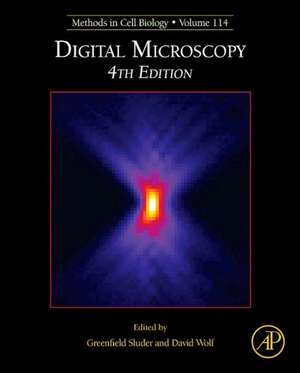
Digital Microscopy
Editat de Greenfield Sluder, David E. Wolfen Limba Engleză Hardback – 25 sep 2013
- Expands coverage to include discussion of confocal microscopy not found in the previous edition
- Includes "traps and pitfalls" as well as laboratory exercises to help illustrate methods
Preț: 624.46 lei
Preț vechi: 901.35 lei
-31% Nou
Puncte Express: 937
Preț estimativ în valută:
119.50€ • 129.76$ • 100.38£
119.50€ • 129.76$ • 100.38£
Carte tipărită la comandă
Livrare economică 16-30 aprilie
Preluare comenzi: 021 569.72.76
Specificații
ISBN-13: 9780124077614
ISBN-10: 0124077617
Pagini: 672
Ilustrații: Illustrationsstrations
Dimensiuni: 191 x 235 x 33 mm
Greutate: 1.36 kg
Ediția:Revised
Editura: ELSEVIER SCIENCE
ISBN-10: 0124077617
Pagini: 672
Ilustrații: Illustrationsstrations
Dimensiuni: 191 x 235 x 33 mm
Greutate: 1.36 kg
Ediția:Revised
Editura: ELSEVIER SCIENCE
Public țintă
Cell and molecular biologists and researchers utilizing digitial microscopy techniques.Cuprins
Microscope Basics.
The Optics of Microscope Image Formation.
Proper Alignment of the Microscope.
Mating Cameras to Microscopes.
Fundamentals of Fluorescence and Fluorescence Microscopy.
Fluorescent Protein Applications in Microscopy.
Live Cell Fluorescence Imaging.
Working with Classic Video.
Practical Aspects of Adjusting Digital Cameras.
Cameras for Digital Microscopy.
A High-Resolution Multimode Digital Microscope System.
Electronic Cameras for Low-Light Microscopy.
Camera Technologies for Low Light Imaging: Overview and Relative Advantages.
Digital Manipulation of Brightfield and Fluorescence Images: Noise Reduction, Contrast Enhancement, and Feature Extraction.
Digital Image Files in Light Microscopy.
High-Resolution Video–Enhanced Differential Interference Contrast Light Microscopy.
Quantitative Analysis of Digital Microscope Images.
Evaluating Optical Aberration Using Fluorescent Microspheres: Methods, Analysis, and Corrective Actions.
Ratio Imaging: Practical Considerations for Measuring Intracellular Ca++ and pH in Living Cells.
Computational Restoration of Fluorescence Images: Noise Reduction, Deconvolution, and Pattern Recognition.
Quantitative Fluorescence Microscopy and Image Deconvolution.
Practical Aspects of Quantitative Confocal Microscopy.
Theoretical Principles and Practical Considerations for Fluorescence Resonance Energy Transfer Microscopy.
Fluorescence Lifetime Imaging Microscopy.
Fluorescence Correlation Spectroscopy: Molecular Complexing in Solution and in Living Cells.
Breaking the Resolution Limit in Light Microscopy.
