Diseases of the Chest: Imaging Diagnosis Based on Pattern Classification
Autor Toshiharu Matsushimaen Limba Engleză Hardback – 21 noi 2006
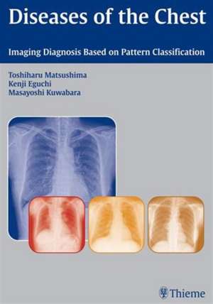
The chest x-ray is the most commonly performed diagnostic x-ray examination. Highly illustrated and with only a minimum of necessary text, this new book takes a highly efficient pattern-based approach to evaluating the chest x-ray. Classes of findings, such as increased radiolucency or alveolar shadow: atelectasis, are used to orient the reader toward the underlying medical problem. Tables help to organize the basic findings so as to be able to arrive at a differential diagnosis.
While the emphasis is on the chest x-ray, a supporting role is played by helpful CT images, schematics and drawings, and photographs of pathologic specimens, where these may be helpful to understand the chest x-ray appearance of the disease.
Chest x-rays are often performed routinely, prior to employment, prior to surgery or during immigration. The examining physician must be in a position to evaluate large numbers of chest x-rays confidently and speedily. This book succeeds admirably in helping the examiner to this end.
Preț: 633.84 lei
Preț vechi: 678.23 lei
-7% Nou
Puncte Express: 951
Preț estimativ în valută:
121.30€ • 126.17$ • 100.14£
121.30€ • 126.17$ • 100.14£
Carte tipărită la comandă
Livrare economică 11-22 aprilie
Preluare comenzi: 021 569.72.76
Specificații
ISBN-13: 9783131435712
ISBN-10: 3131435712
Pagini: 183
Ilustrații: 601
Dimensiuni: 220 x 306 x 18 mm
Greutate: 0.91 kg
Ediția:1st edition
Editura: Thieme
Colecția Thieme
ISBN-10: 3131435712
Pagini: 183
Ilustrații: 601
Dimensiuni: 220 x 306 x 18 mm
Greutate: 0.91 kg
Ediția:1st edition
Editura: Thieme
Colecția Thieme
Recenzii
Well organized...The quality of the radiologic images in this text is excellent, with a generous quantity of accompanying color diagrams, charts, bronchoscopy photographs, and gross and microscopic specimen photographs.--RadiologyAn easy-to-follow format...provides a rational and useful approach for evaluating chest radiographs...the abundant images are excellent and the image quality is superior...provides an excellent review for practitioners with advanced training.--Doody's Book Reviews
Textul de pe ultima copertă
The chest x-ray is the most commonly performed diagnostic x-ray examination. Highly illustrated and with only a minimum of necessary text, this new book takes a highly efficient pattern-based approach to evaluating the chest x-ray. Classes of findings, such as increased radiolucency or alveolar shadow: atelectasis, are used to orient the reader toward the underlying medical problem. Tables help to organize the basic findings so as to be able to arrive at a differential diagnosis.
While the emphasis is on the chest x-ray, a supporting role is played by helpful CT images, schematics and drawings, and photographs of pathologic specimens, where these may be helpful to understand the chest x-ray appearance of the disease.
Chest x-rays are often performed routinely, prior to employment, prior to surgery or during immigration. The examining physician must be in a position to evaluate large numbers of chest x-rays confidently and speedily. This book succeeds admirably in helping the examiner to this end.
While the emphasis is on the chest x-ray, a supporting role is played by helpful CT images, schematics and drawings, and photographs of pathologic specimens, where these may be helpful to understand the chest x-ray appearance of the disease.
Chest x-rays are often performed routinely, prior to employment, prior to surgery or during immigration. The examining physician must be in a position to evaluate large numbers of chest x-rays confidently and speedily. This book succeeds admirably in helping the examiner to this end.
