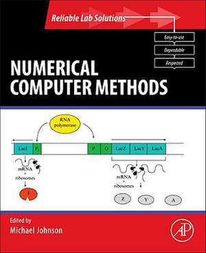
Essential Numerical Computer Methods: Reliable Lab Solutions
Editat de Michael L. Johnsonen Limba Engleză Paperback – 24 noi 2010
A general perception exists that the only applications of computers and computer methods in biological and biomedical research are either basic statistical analysis or the searching of DNA sequence data bases. While these are important applications they only scratch the surface of the current and potential applications of computers and computer methods in biomedical research. The various chapters within this volume include a wide variety of applications that extend far beyond this limited perception. As part of the Reliable Lab Solutions series, Essential Numerical Computer Methods brings together chapters from volumes 210, 240, 321, 383, 384, 454, and 467 of Methods in Enzymology. These chapters provide a general progression from basic numerical methods to more specific biochemical and biomedical applications.
- The various chapters within this volume include a wide variety of applications that extend far beyond this limited perception
- As part of the Reliable Lab Solutions series, Essential Numerical Computer Methods brings together chapters from volumes 210, 240, 321, 383, 384, 454, and 467 of Methods in Enzymology
- These chapters provide a general progression from basic numerical methods to more specific biochemical and biomedical applications
Preț: 487.44 lei
Preț vechi: 633.05 lei
-23% Nou
Puncte Express: 731
Preț estimativ în valută:
93.27€ • 97.64$ • 77.18£
93.27€ • 97.64$ • 77.18£
Carte tipărită la comandă
Livrare economică 05-19 aprilie
Preluare comenzi: 021 569.72.76
Specificații
ISBN-13: 9780123849977
ISBN-10: 0123849977
Pagini: 616
Ilustrații: 1
Dimensiuni: 191 x 235 x 28 mm
Greutate: 1.07 kg
Editura: ELSEVIER SCIENCE
Seria Reliable Lab Solutions
ISBN-10: 0123849977
Pagini: 616
Ilustrații: 1
Dimensiuni: 191 x 235 x 28 mm
Greutate: 1.07 kg
Editura: ELSEVIER SCIENCE
Seria Reliable Lab Solutions
Public țintă
Biochemists, biophysicists, physical chemists, molecular biologists, cell biologistsCuprins
Part
I:
Fluorochromes/General
Techniques1.
Principles
of
confocal
microscopy
2.
Protein
labeling
with
fluorescent
probes
3.
Cytometry
of
fluorescence
resonance
energy
transfer
4.
The
rainbow
of
fluorescent
proteins
5.
Labeling
cellular
targets
with
semiconductor
quantum
dot
conjugates
6.
High-gradient
magnetic
cell
sorting
7.
Multiplexed
microsphere
assays
for
protein
and
DNA
binding
reactions
8.
Biohazard
sorting
9.
Guidelines
for
the
presentation
of
flow
cytometric
data
Part II: Cellular DNA Content Analysis10. VinDetergent and proteolytic enzyme-based techniques for nuclear isolation and DNA content analysis 11. Rapid DNA content analysis 12. DNA analysis from paraffin-embedded blocks 13. Flow cytometry and sorting of plant protoplasts and cells 14. DNA content histogram and cell cycle analysis 15. Simultaneous analysis of cellular RNA and DNA content
Part III. Cell Proliferation and Death Assays16. Immunochemical quantitation of bromodeoxyuridine: application to cell cycle kinetics 17. Cell cycle kinetics estimated by analysis of bromodeoxyuridine incorporation 18. Flow cytometric analysis of cell division history using dilution of carboxyfluorescein diacetate succinimidyl ester, a stably integrated fluorescent probe 19. Antibodies against the Ki-67 protein: Assessment of the growth fraction and tools for cell cycle analysis 20. Detection of DNA damage in individual cells by analysis of histone H2AX phosphorylation 21. Assays of cell viability: Discrimination of cells dying by apoptosis 22. Difficulties and pitfalls in analysis of apoptosis
Part IV: Cell Surface Immunophenotyping23. Cell preparation for the identification of leukocytes 24. Multicolor immunophenotyping: Human immune system hematopoiesis 25. Differential diagnosis of T-cell lymphoproliferative disorders by flow cytometry multicolor immunophenotyping. Correlation with morphology 26. B-cell immunophenotyping
Part V: Cytogenetics/Chromatin Structure27. Telomere length measurements using in situ hybridization 28. Sperm chromatin structure assay: DNA denaturability
Part VI: Cell Physiology Assays29. Cell membrane potential analysis 30. Measurement of intracellular pH 31. Intracellular ionized calcium 32. Oxidative product formation analysis by flow cytometry 33. Phagocyte function 34. Analysis of RNA synthesis by cytometry 35. Analysis of mitochondria by flow cytometry 36. Analysis of platelets by flow cytometry
Part VII: Detection of Microorganisms and Pathogens37. Detection of specific microorganisms in environmental samples using flow cytometry 38. Flow cytometric analysis of microorganisms 39. Flow cytometry of malaria detection
