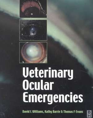
Handbook of Veterinary Ocular Emergencies
Autor David A. Williams, Kathy Barrie Editat de Thomas Ffrangcon Evansen Limba Engleză Paperback – 8 apr 2002
Preț: 528.96 lei
Preț vechi: 556.79 lei
-5% Nou
Puncte Express: 793
Preț estimativ în valută:
101.23€ • 105.29$ • 83.57£
101.23€ • 105.29$ • 83.57£
Carte disponibilă
Livrare economică 24 martie-07 aprilie
Preluare comenzi: 021 569.72.76
Specificații
ISBN-13: 9780750635608
ISBN-10: 0750635606
Pagini: 128
Ilustrații: 40 ills.
Dimensiuni: 186 x 244 x 7 mm
Greutate: 0.29 kg
Editura: Elsevier
ISBN-10: 0750635606
Pagini: 128
Ilustrații: 40 ills.
Dimensiuni: 186 x 244 x 7 mm
Greutate: 0.29 kg
Editura: Elsevier
Public țintă
Vets in practice worldwide; veterinary students; veterinary nurses and techniciansCuprins
FOREWORD
INTRODUCTORY CHAPTERS
CHAPTER 1: INTRODUCTION
1.1 How to use this book
1.2 Performing an ocular examination in an emergency situation
1.3 Recording observations made in an ocular emergency
1.4 Equipment and aids required to deal with the ocular emergency
1.5 Some preliminary notes on treatment of ocular infections
1.6 Analgesia in ocular emergencies
1.7 Dealing with ocular emergencies in horses and ruminants
1.7.1 Techniques facilitating large animal ocular examination
1.7.2Techniques facilitating large animal ocular therapeutics
CHAPTER 2: A problem orientated approach
2.1: The red eye
2.2 The painful eye
2.3 The white eye
2.4 The suddenly blind eye
2.5 Ocular lesions in systemic disease
DIAGNOSIS AND TREATMENT OF OCULAR EMERGENCIES
CHAPTER 3: ADNEXA AND ORBIT
3.1: Lid laceration
3.2 Conjunctivitis
3.3 Conjunctival foreign body
3.4 Acute keratoconjunctivitis sicca
3.5 Orbital cellulitis
3.6 Orbital space occupying lesion
CHAPTER 4: GLOBE
4.1: Blunt trauma to the globe
4.2: Globe prolapse
4.3: Penetrating globe injury
CHAPTER 5: CORNEA
5.1: Corneal ulceration
5.1.1:Is an ulcer present? - the use of ophthalmic stains
5.1.2: Three key questions regarding any corneal ulcer
5.1.2.1 Ulcer depth
5.1.2.2 Ulcer healing
5.1.2.3 The cause of the ulcer
5.2 Dealing with different ulcers
5.2.1The simple healing superficial ulcer
5.2.2The recurrent or persistent non-healing superficial ulcer
5.2.3Ulceration secondary to bullous keratopathy
5.2.4Partial thickness stromal ulceration
5.2.5 Near-penetrating ulcers, descemetocoeles and penetrating ulcers
5.2.6.1 The melting ulcer: diagnosis
5.2.6.2 The melting ulcer: diagnosis
5.3 Corneoscleral laceration
5.3.1Defining the extent of a corneal laceration
5.3.2Defining involvement of other ocular structures
5.3.3Repairing a simple non-penetrating corneal laceration
5.3.4Repairing a simple perforating corneal laceration
5.3.5 Repairing a corneal laceration complicated by iris inclusion
5.4 Corneal foreign bodies
5.4.1 Recognising a corneal foreign body
5.4.2 Dealing with a non-perforating corneal foreign body
5.4.3 Dealing with a fully penetrating corneal foreign body
5.5 Antibiotics and mydriatic cycloplegia in corneal emergencies
CHAPTER 6: IRIS
6.1 Iritis
6.1.1Diagnosis: clinical signs
6.1.2 Diagnosis: diagnostic tests
6.1.3 Treatment: pain relief
6.1.4 Treatment: anti-inflammatory medication
6.1.5 Treatment: reducing miosis and preventing synechia formation
6.2Change in iris appearance
CHAPTER 7: GLAUCOMA
7.1 Diagnosis: clinical signs
7.2 Diagnosis: diagnostic tests
7.3 Treatment: immediate systemic hypotensive therapy
7.4 Treatment: long-term reduction of IOP
7.5 Treatment: neuroprotection
CHAPTER 8: LENS
8.1 Lens luxation
8.2 Diabetic cataract
8.3 Lens capsule rupture and phacoanaphylactic uveitis
CHAPTER 9: RETINA AND VITREOUS
9.1 Retinal detachment
9.1.1 Examination of the animal with a retinal detachment
9.1.2 Treatment of retinal detachment secondary to hypertension
9.1.3 Treatment of retinal detachment in posterior uveitis
9.1.4 Treatment of idiopathic retinal detachment
9.2 Sudden acquired retinal degeneration (SARD)
CHAPTER 10: OPTIC NERVE
10.1Optic neuritis
10.2Central blindness
CHAPTER 11: CONCLUSIONS
APPENDIX:
Section 1: Diagnostic methods used in veterinary ophthalmology
Section 2: Ocular Dictionary
Section 3: Ocular Formulary


