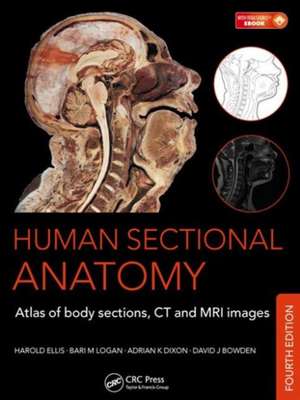
Human Sectional Anatomy: Atlas of Body Sections, CT and MRI Images, Fourth Edition
Autor Adrian K. Dixon, David J. Bowden, Harold Ellis, Bari M. Loganen Limba Engleză Hardback – 24 apr 2015
The superb full-colour cadaver sections are compared with CT and MRI images, with accompanying, labelled, line diagrams. Many of the radiological images have been replaced with new examples for this latest edition, captured using the most up-to date imaging technologies to ensure excellent visualization of the anatomy. The photographic material is enhanced by useful notes with details of important anatomical and radiological features.
Beautifully presented in a generous format, Human Sectional Anatomy continues to be an invaluable resource for all radiologists, radiographers, surgeons and medics, in training and in practice, and an essential component of departmental and general medical library collections.
Preț: 1147.29 lei
Preț vechi: 1380.54 lei
-17% Nou
Puncte Express: 1721
Preț estimativ în valută:
219.55€ • 227.83$ • 183.51£
219.55€ • 227.83$ • 183.51£
Comandă specială
Livrare economică 24 februarie-10 martie
Doresc să fiu notificat când acest titlu va fi disponibil:
Se trimite...
Preluare comenzi: 021 569.72.76
Specificații
ISBN-13: 9781498703604
ISBN-10: 1498703607
Pagini: 288
Ilustrații: 428 colour illustrations
Dimensiuni: 250 x 330 x 23 mm
Greutate: 1.78 kg
Ediția:Revised
Editura: CRC Press
Colecția CRC Press
ISBN-10: 1498703607
Pagini: 288
Ilustrații: 428 colour illustrations
Dimensiuni: 250 x 330 x 23 mm
Greutate: 1.78 kg
Ediția:Revised
Editura: CRC Press
Colecția CRC Press
Public țintă
Professional ReferenceCuprins
BRAIN. Superficial dissection. Selected images. HEAD. Axial sections (male). Selected images: axial MRI. Selected images: temporal bone/inner ear: axial CT. Coronal sections (female). Sagittal sections (male). NECK. Axial sections (female). Sagittal sections (male). THORAX. Axial sections (male). Axial sections (female). Selected images: heart. Selected images: mediastinum: axial CT. Selected images: coronal MRI. Selected images: coronal chest CT. Selected images: coeliac and great vessels. ABDOMEN. Axial sections (male). Axial sections (female). Selected images: lumbar spine: axial CT. Selected images: lumbar spine: coronal MRI. Selected images: lumbar spine: sagittal MRI. PELVIS. Axial sections (male). Selected images: coronal MRI (male). Axial sections (female). Selected images: axial MRI (female). Selected images: coronal MRI (female). Selected images: sagittal MRI (female). Selected images: colon. Selected images: coronal abdominal CT. LOWER LIMB. Hip: coronal sections (female). Selected images: pelvic girdle. Thigh: axial sections (male). Knee: axial sections (male). Knee: coronal sections (male). Knee: sagittal sections (female). Leg: axial sections (male). Ankle: axial sections (male). Ankle: coronal sections (female). Foot: sagittal sections (male). Foot: coronal sections (male). UPPER LIMB. Shoulder: axial sections (female). Shoulder: coronal sections (male). Arm: axial sections (male). Elbow: axial sections (male). Elbow: coronal sections (female). Forearm: axial sections (male). Wrist: axial sections (male). Hand: coronal sections (female). Hand: sagittal sections (female). Hand: axial sections (male). Selected images: shoulder girdle.
Notă biografică
Harold Ellis, CBE, MA, DM, MCH, FRCS, FRCOG, professor, Applied Clinical Anatomy Group, Applied Biomedical Research, Guy’s Hospital, London, UK
Bari M. Logan, MA, FMA, Hon MBIE, MAMAA, formerly university prosector, Department of Anatomy, University of Cambridge, UK; and formerly prosector, Department of Anatomy, The Royal College of Surgeons of England, London, UK
Adrian K. Dixon, MD, FRCP, FRCR, FRCS, FMedSci, emeritus professor, Department of Radiology, University of Cambridge, UK; honorary consultant radiologist, Addenbrooke’s Hospital, Cambridge, UK; and master, Peterhouse, University of Cambridge, UK
David J. Bowden, MA, VetMB, MB BChir, FRCR, abdominal imaging fellow, Department of Medical Imaging, Sunnybrook Health Sciences Centre, Toronto, Canada; and formerlyteaching bye-fellow, Christ’s College, University of Cambridge, UK
Descriere
This book set new standards for the quality of cadaver sections and accompanying radiological images. Now in its fourth edition, this unsurpassed quality remains and is further enhanced by additional new material. The superb full-colour cadaver sections are compared with CT and MRI images, with accompanying, labelled, line diagrams. Many of the radiological images have been replaced with new examples for this latest edition, captured using the most up-to date imaging technologies to ensure excellent visualization of the anatomy. The photographic material is enhanced by useful notes with details of important anatomical and radiological features.
