Cantitate/Preț
Produs

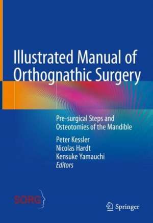
Illustrated Manual of Orthognathic Surgery: Osteotomies of the Mandible

Editat de Peter Kessler, Nicolas Hardt, Kensuke Yamauchi
en Limba Engleză Hardback – 15 mar 2024
This first volume in a multi-volume series considers the gains in information and knowledge that have resulted from preoperative and postoperative 3D imaging using new radiologic protocols in maxillofacial surgery, with the corresponding consequences for the surgeon. It contrasts the established standard techniques of orthognathic oral and maxillofacial surgery with new considerations and insights based on years of experience and analysis of clinical activity in this subspecialty of oral and maxillofacial surgery.
The book is deliberately structured so that the clinical situation is juxtaposed with a graphic representation for better understanding, which is intended to point out special situations that can in turn positively influence the surgical planning and execution of the procedure in order to avoid undesirable results in individual cases. Graphic representations illustrate the three-dimensionality of the complex splitting technique of the mandible where text or radiographs are insufficient.

A corresponding book that combines clinical anatomical situations with pre- and postoperative preparation and clear explanation does not yet exist. It is aimed at a wide readership of students and professionals in oral and maxillofacial surgery, orthodontics, plastic and craniofacial surgery, and otolaryngology.
Citește tot Restrânge

Preț: 104705 lei

Preț vechi: 110215 lei
-5% Nou

Puncte Express: 1571

Preț estimativ în valută:
20038 20815$ 16645£

Carte disponibilă

Livrare economică 13-27 ianuarie 25
Livrare express 27 decembrie 24 - 02 ianuarie 25 pentru 5565 lei

Preluare comenzi: 021 569.72.76

Specificații

ISBN-13: 9783031069772
ISBN-10: 3031069773
Pagini: 360
Ilustrații: X, 360 p. 289 illus., 274 illus. in color.
Dimensiuni: 178 x 254 x 27 mm
Greutate: 0.98 kg
Ediția:2024
Editura: Springer International Publishing
Colecția Springer
Locul publicării:Cham, Switzerland

Cuprins

Introduction to Orthopaedic Surgery in the Mandible.- Ramus-Split-Osteotomies / Bilateral Sagittal Split Osteotomies (BSSO): General Planning.- Ramus-Split-Osteotomies (BSSO).- Surgical Principles.- Mandibular Deficiency.- Surgical Technique.- BSSO.- Mandibular Excess.- Surgical Technique.- BSSO.- Asymmetries, Vertical and Horizontal rotation, mandibular Flaring.- Surgical Techniques.- Mandibular excess.- Class III setback.- Surgical Technique-IVRO.- Alveolar Segment Osteotomies.- Chin Osteotomies.- The Temporomandibular Joint.


Notă biografică

Peter A. Kessler is Professor and Head of the Department of Cranio-Maxillofacial Surgery, Maastricht University Medical Center (MUMC), Maastricht, The Netherlands. Dr. Kessler completed his studies in dentistry and medicine at the Friedrich-Alexander-University of Erlangen-Nuremberg in 1986 and 1994 respectively (Prof. Dr. Emil Steinhäuser), where he habilitated in 2001. He qualified as Specialist in Oral and Maxillofacial Surgery in 1999 under Prof. Dr. F.W. Neukam and holds a qualification in plastic surgery (2003). From 1999 to 2001 he worked as a specialist at the Department of Oral and Maxillofacial Surgery, Friedrich-Alexander University Erlangen-Nuremberg, Erlangen (Prof. Dr. Neukam); he then became Senior Lecturer and Vice-Director at the clinic before taking up his current position in Maastricht in 2007.

 
Nicolas P. Hardt was Professor and Head of the Clinic for Cranio-Maxillofacial Surgery, Kantonsspital, Lucerne (LUKS), Switzerland from 1974 until 2007. Dr. Hardt graduated in Dentistry and Medicine at the University of Bonn, Germany, and subsequently completed a residency at the Clinic for Cranio-Maxillofacial Surgery in Bonn (Prof. Dr. Eberhard Krüger). In 1970 he was appointed Senior Assistant in the Clinic for Cranio-Maxillofacial Surgery at the University of Berne in Switzerland (Prof. Dr. Otto Neuner). In 1973 he was visiting maxillo-facial Surgeon in the Centre for Cranio-Maxillofacial Surgery / Queen Mary’s-Hospital - Roehampton/London (Sir Norman Rowe). He then qualified by Prof. Dr. Emil Steinhäuser (Erlangen/Nürnberg) as a Specialist for Cranio-Maxillofacial Surgery in Switzerland (1974) and Germany (1975) and the EU (1979) and holds a qualification in Plastic Surgery (1980). Dr. Hardt completed his Habilitation by Prof. Dr. Hugo Obwegeser (University Clinic for Cranio-Maxillofacial Surgery of Zürich) in 1981 and became a Full Professor at the University of Zurich in 1990.
 
Kensuke Yamauchi is Associate Professor at the Department of Oral and Maxillofacial Surgery, Tohoku University, Sendai, Japan (Head: Prof. Tetsu Takahashi). Dr. Yamauchi graduated in Dentistry at Tohoku University in 2001 and completed a residency at the Kagawa Prefectural Central Hospital in Kagawa (Dr. Masaharu Mitsugi). He was appointed assistant professor at the Department of Oral and Maxillofacial Reconstructive Surgery of Kyushu Dental College (Prof. Tetsu Takahashi) until 2012. He also visited the Department of Cranio-Maxillofacial Surgery, Maastricht University Medical Center (Prof. Dr. Peter Kessler) from 2011 to 2012. He moved to the Department of Oral and Maxillofacial Surgery, Tohoku University (Prof. Tetsu Takahashi) in 2012, and became a Lecturer and Vice-Director of Dental Implant Center in 2013 and taking up his current position in Tohoku University in 2017.

Textul de pe ultima copertă

This first volume in a multi-volume series considers the gains in information and knowledge that have resulted from preoperative and postoperative 3D imaging using new radiologic protocols in maxillofacial surgery, with the corresponding consequences for the surgeon. It contrasts the established standard techniques of orthognathic oral and maxillofacial surgery with new considerations and insights based on years of experience and analysis of clinical activity in this subspecialty of oral and maxillofacial surgery.
 
The book is deliberately structured so that the clinical situation is juxtaposed with a graphic representation for better understanding, which is intended to point out special situations that can in turn positively influence the surgical planning and execution of the procedure in order to avoid undesirable results in individual cases. Graphic representations illustrate the three-dimensionality of the complex splitting technique of the mandible where text or radiographs are insufficient.
 
A corresponding book that combines clinical anatomical situations with pre- and postoperative preparation and clear explanation does not yet exist. It is aimed at a wide readership of students and professionals in oral and maxillofacial surgery, orthodontics, plastic and craniofacial surgery, and otolaryngology.

Caracteristici

Ensures fast access to information and step-by-step learning
Updates readers of the relationship between clinical situation, radiological workup, and post-op analysis.
Deepens readers comprehension of surgical problems

Descriere

This first volume in a multi-volume series considers the gains in information and knowledge that have resulted from preoperative and postoperative 3D imaging using new radiologic protocols in maxillofacial surgery, with the corresponding consequences for the surgeon. It contrasts the established standard techniques of orthognathic oral and maxillofacial surgery with new considerations and insights based on years of experience and analysis of clinical activity in this subspecialty of oral and maxillofacial surgery.

 
The book is deliberately structured so that the clinical situation is juxtaposed with a graphic representation for better understanding, which is intended to point out special situations that can in turn positively influence the surgical planning and execution of the procedure in order to avoid undesirable results in individual cases. Graphic representations illustrate the three-dimensionality of the complex splitting technique of the mandible where text or radiographs are insufficient.
 
A corresponding book that combines clinical anatomical situations with pre- and postoperative preparation and clear explanation does not yet exist. It is aimed at a wide readership of students and professionals in oral and maxillofacial surgery, orthodontics, plastic and craniofacial surgery, and otolaryngology.