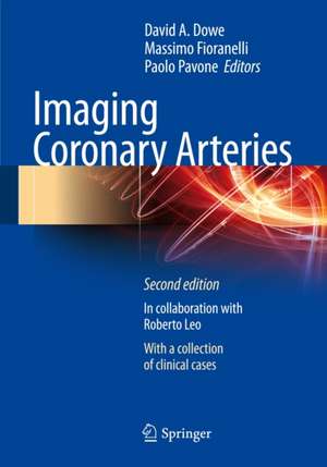
Imaging Coronary Arteries
Editat de David A. Dowe, Massimo Fioranelli, Paolo Pavoneen Limba Engleză Paperback – 6 dec 2012
Preț: 728.16 lei
Preț vechi: 766.49 lei
-5% Nou
Puncte Express: 1092
Preț estimativ în valută:
139.38€ • 151.45$ • 117.15£
139.38€ • 151.45$ • 117.15£
Carte disponibilă
Livrare economică 31 martie-14 aprilie
Preluare comenzi: 021 569.72.76
Specificații
ISBN-13: 9788847026810
ISBN-10: 8847026814
Pagini: 261
Ilustrații: XII, 261 p.
Dimensiuni: 178 x 254 x 15 mm
Greutate: 0.73 kg
Ediția:2nd ed. 2013
Editura: Springer
Colecția Springer
Locul publicării:Milano, Italy
ISBN-10: 8847026814
Pagini: 261
Ilustrații: XII, 261 p.
Dimensiuni: 178 x 254 x 15 mm
Greutate: 0.73 kg
Ediția:2nd ed. 2013
Editura: Springer
Colecția Springer
Locul publicării:Milano, Italy
Public țintă
Professional/practitionerCuprins
1 Clinical Anatomy of the Coronary Circulation.- 2 Basic Techniques in the Acquisition of Cardiac Images with CT.- 3 Examination of the Coronary Arteries.- 4 Image Reconstruction.- 5 Coronary Pathophysiology.- 6 Clinical Classification of Coronary Artery Disease: Who Should Be Treated?.- 7 Intravascular Ultrasound: From Gray-Scale to Virtual Histology.- 8 Identification and Characterization of the Atherosclerotic Plaque Using Coronary CT Angiography.- 9 Coronary CT Angiography: Evaluation of Stenosis and Occlusion.- 10 Current Strategies in Cardiac Surgery.- 11 Coronary CT Angiography: Evaluation of Coronary Artery Bypass Grafts.- 12 Coronary Stents.- 13 CT Angiography of Coronary Stents.- 14 X-Ray Exposure in Coronary CT Angiography.- 15 Optical Coherence Tomography in the Cathlab.- 16 Triple Rule Out: The Use of Cardiac CT in the Emergency Room.- 17 Contraindications to Coronary CT Angiography.- 18 Prognostic Value of Coronary CT.- 19 Clinical Cases.
Textul de pe ultima copertă
Cardiovascular diseases are the leading cause of death in Western countries. In non-fatal cases, they are associated with a decreased quality of life as well as a substantial economic burden to society. Most sudden cardiac events are related to the complications of a non-stenosing marginal plaque. For this reason, the ability to properly identify the atherosclerotic plaque with rapid, non-invasive techniques is of utmost clinical interest in therapeutic planning. CT produces high-quality images of the coronary arteries, in addition to defining their location and the extent of the atherosclerotic involvement. Proper knowledge of the equipment, adequate preparation of the patient, and accurate evaluation of the images are essential to obtaining a consistent clinical diagnosis in every case. This new edition is enriched with dedicated chapters on intravascular ultrasound (IVUS), catheter angiography, and nuclear imaging, with limited discussions of theoretical techniques such as optical coherence tomography (OCT) and magnetic resonance imaging (MRI). Brief chapters will also be included that discuss percutaneous coronary artery procedures and coronary artery bypass surgery. Indications of when to use CCTA will be discussed, with comparison of these imaging techniques to each other. This volume provides general practitioners and cardiologists with a basic understanding of the imaging techniques. For radiologists with no direct experience in cardiac imaging, the book serves as an important source of information on coronary pathophysiology and anatomy.
Caracteristici
New dedicated chapters on intravascular ultrasound (IVUS), catheter angiography, and nuclear imaging, with some discussions on theoretical techniques such as optical coherence tomography (OCT) and magnetic resonance imaging (MRI) More than 70 new clinical cases
