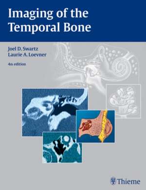
Imaging of the Temporal Bone
Autor Joel D. Swartz, Laurie A. Loevneren Limba Engleză Hardback – 2 noi 2008
Praise for this book:
This book is highly recommended and should find its way onto the library shelf of every neuroradiology section. - American Journal of Neuroradiology
Authoritative and lavishly illustrated, this best-selling reference returns in a fourth edition with comprehensive coverage of the current imaging strategies for the evaluation of disease processes affecting the temporal bone and its intricate anatomy. New in this edition is a highly practical how-to chapter that presents imaging modalities and technical parameters for CT and MRI as well as an overview of the role of plain film radiography, ultrasound, PET, and PET/CT. The chapter then addresses major clinical indications, providing step-by-step descriptions of how to protocol each case, how to interpret the studies, and how to report findings. The remaining chapters thoroughly cover specific anatomic areas of the temporal bone separately. Each chapter places special emphasis on gaining a solid foundation of the normal anatomy and anatomic variations. It then discusses imaging protocols and image evaluation for specific clinical problems.
Highlights:
- Practical discussion of standard techniques, protocols, and special considerations for imaging using CT and MRI
- In-depth coverage of both common and rare conditions
- Clinical insights from international authorities in the field
- More than 1,500 high-quality illustrations and images, including CT, MRI, and vascular images using CTA, MRA, and conventional catheter angiography
Preț: 905.03 lei
Preț vechi: 1235.84 lei
-27% Nou
Puncte Express: 1358
Preț estimativ în valută:
173.17€ • 181.30$ • 143.29£
173.17€ • 181.30$ • 143.29£
Carte indisponibilă temporar
Doresc să fiu notificat când acest titlu va fi disponibil:
Se trimite...
Preluare comenzi: 021 569.72.76
Specificații
ISBN-13: 9781588903457
ISBN-10: 1588903451
Pagini: 604
Ilustrații: 1506
Dimensiuni: 225 x 286 x 31 mm
Greutate: 1.9 kg
Ediția:Revizuită
Editura: MM – Thieme
ISBN-10: 1588903451
Pagini: 604
Ilustrații: 1506
Dimensiuni: 225 x 286 x 31 mm
Greutate: 1.9 kg
Ediția:Revizuită
Editura: MM – Thieme
Recenzii
Comprehensive coverage of the current imaging strategies for the evaluation of disease processes affecting the temporal bone. -- The Neuroradiology JournalThe latest edition of one of the great landmark treatises in neuroradiology...a reference textbook that should be part of any radiologist's library...has been extensively rewritten...the illustrations have been extensively updated, including cutting-edge, state-of-the-art, and painstakingly labeled CT and MRI images...useful...improves on the previous masterpiece.--American Journal of Roentgenology[The authors] have expanded the next by nearly 100 pages...and, in doing so, have been able to incorporate more images and increasingly important details in the text...an in-depth evaluation...This book is highly recommended and should find its way onto the library shelf of every neuroradiology section. For those who interpret many temporal bone studies, a personal copy is warranted.--American Journal of NeuroradiologyA satisfying resource. This new edition contain exceptional imaging techniques, reporting information, and, most of all, elaborately illustrated anatomical images accompanied by examples of real CT and MR scans. Its attention to protocol detail and depiction of anatomy distinguish the book from many similarly themed texts...With the amount of concise and comprehensive information contained in the fourth edition, this book is surprisingly easy to read and understand. This valuable teaching and resource tool would be well-received in any reading room.--Advance for Imaging & Radiation OncologyA concise textbook...the text is very well organized, well focused, and, as one would expect with any imaging text, replete with exemplary images thoroughly covering...this complex anatomic region. Beginning with the table of contents, one sees that the text is efficiently organized to take readers into the subject matter starting laterally at the external auditory canal and ending medially at the internal auditory canal and nearby intracranial contents...strongly recommend[ed].--Otology & NeurotologyA comprehensive textbook...A great deal of attention has been placed on providing thorough details in charts, images and drawings. The information flows well, which makes it easier to understand...The strong points of this textbook are the in-depth information and organization...an extensive and detailed study of the temporal bone.--Radiologic TechnologyExtensively revised to demonstrate a decade's advances in imaging. This text remains more than a simple atlas, although, being lavishly illustrated with over 1,500 images, an atlas it undoubtedly is. The content is well updated...Very thought provoking but extremely complex...essential in any ENT department aspiring to train: it represents excellent value.--The Journal of Laryngology & OtologyPraise for previous editions:The writing is lucid, illustrations are superb and detailed, and line drawings are excellent...This book is a necessity... --RadiologyA valuable resource for radiology residents, fellows and practicing radiologists...The authors have done an excellent job of compiling MR images that show typical disease process...A high-quality product. --American Journal of RoentgenologyExcellent...well-written...This text should be read cover-to-cover by all trainees and practicing neuroradiologists. Moreover, it should be readily available in every departments library.--American Journal of NeuroradiologyComprehensive, well-written, well-illustrated...Otologists, lateral skull base surgeons, and radiologists should find this text to be a valuable addition to their reference collection.--Head and Neck
Notă biografică
Professor of Radiology and Otorhinolaryngology: Head and Neck Surgery, and Neurosurgery, University of Pennsylvania Medical Center, Philadelphia, PA, USA
Textul de pe ultima copertă
Praise for this book:
This book is highly recommended and should find its way onto the library shelf of every neuroradiology section. - American Journal of Neuroradiology
Authoritative and lavishly illustrated, this best-selling reference returns in a fourth edition with comprehensive coverage of the current imaging strategies for the evaluation of disease processes affecting the temporal bone and its intricate anatomy. New in this edition is a highly practical how-to chapter that presents imaging modalities and technical parameters for CT and MRI as well as an overview of the role of plain film radiography, ultrasound, PET, and PET/CT. The chapter then addresses major clinical indications, providing step-by-step descriptions of how to protocol each case, how to interpret the studies, and how to report findings. The remaining chapters thoroughly cover specific anatomic areas of the temporal bone separately. Each chapter places special emphasis on gaining a solid foundation of the normal anatomy and anatomic variations. It then discusses imaging protocols and image evaluation for specific clinical problems.
Highlights:
This book is highly recommended and should find its way onto the library shelf of every neuroradiology section. - American Journal of Neuroradiology
Authoritative and lavishly illustrated, this best-selling reference returns in a fourth edition with comprehensive coverage of the current imaging strategies for the evaluation of disease processes affecting the temporal bone and its intricate anatomy. New in this edition is a highly practical how-to chapter that presents imaging modalities and technical parameters for CT and MRI as well as an overview of the role of plain film radiography, ultrasound, PET, and PET/CT. The chapter then addresses major clinical indications, providing step-by-step descriptions of how to protocol each case, how to interpret the studies, and how to report findings. The remaining chapters thoroughly cover specific anatomic areas of the temporal bone separately. Each chapter places special emphasis on gaining a solid foundation of the normal anatomy and anatomic variations. It then discusses imaging protocols and image evaluation for specific clinical problems.
Highlights:
- Practical discussion of standard techniques, protocols, and special considerations for imaging using CT and MRI
- In-depth coverage of both common and rare conditions
- Clinical insights from international authorities in the field
- More than 1,500 high-quality illustrations and images, including CT, MRI, and vascular images using CTA, MRA, and conventional catheter angiography
Descriere
Praise for this book:
This book is highly recommended and should find its way onto the library shelf of every neuroradiology section. - American Journal of Neuroradiology
Authoritative and lavishly illustrated, this best-selling reference returns in a fourth edition with comprehensive coverage of the current imaging strategies for the evaluation of disease processes affecting the temporal bone and its intricate anatomy. New in this edition is a highly practical how-to chapter that presents imaging modalities and technical parameters for CT and MRI as well as an overview of the role of plain film radiography, ultrasound, PET, and PET/CT. The chapter then addresses major clinical indications, providing step-by-step descriptions of how to protocol each case, how to interpret the studies, and how to report findings. The remaining chapters thoroughly cover specific anatomic areas of the temporal bone separately. Each chapter places special emphasis on gaining a solid foundation of the normal anatomy and anatomic variations. It then discusses imaging protocols and image evaluation for specific clinical problems.
Highlights:
This book is highly recommended and should find its way onto the library shelf of every neuroradiology section. - American Journal of Neuroradiology
Authoritative and lavishly illustrated, this best-selling reference returns in a fourth edition with comprehensive coverage of the current imaging strategies for the evaluation of disease processes affecting the temporal bone and its intricate anatomy. New in this edition is a highly practical how-to chapter that presents imaging modalities and technical parameters for CT and MRI as well as an overview of the role of plain film radiography, ultrasound, PET, and PET/CT. The chapter then addresses major clinical indications, providing step-by-step descriptions of how to protocol each case, how to interpret the studies, and how to report findings. The remaining chapters thoroughly cover specific anatomic areas of the temporal bone separately. Each chapter places special emphasis on gaining a solid foundation of the normal anatomy and anatomic variations. It then discusses imaging protocols and image evaluation for specific clinical problems.
Highlights:
- Practical discussion of standard techniques, protocols, and special considerations for imaging using CT and MRI
- In-depth coverage of both common and rare conditions
- Clinical insights from international authorities in the field
- More than 1,500 high-quality illustrations and images, including CT, MRI, and vascular images using CTA, MRA, and conventional catheter angiography
