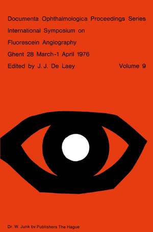
International Symposium on Fluorescein Angiography Ghent 28 March-1 April 1976: Documenta Ophthalmologica Proceedings Series, cartea 9
Editat de J.J. de Laeyen Limba Engleză Paperback – 30 iun 1976
Din seria Documenta Ophthalmologica Proceedings Series
- 5%
 Preț: 358.48 lei
 - 5%
 Preț: 378.97 lei
 - 5%
 Preț: 1405.29 lei
 - 5%
 Preț: 360.34 lei
 - 5%
 Preț: 375.34 lei
 - 5%
 Preț: 1427.79 lei
 - 5%
 Preț: 372.03 lei
 - 5%
 Preț: 1419.39 lei
 - 5%
 Preț: 1427.79 lei
 - 5%
 Preț: 373.47 lei
 - 5%
 Preț: 379.89 lei
 - 5%
 Preț: 352.84 lei
 - 5%
 Preț: 366.35 lei
 - 5%
 Preț: 383.72 lei
 - 5%
 Preț: 1424.52 lei
 - 5%
 Preț: 368.93 lei
 - 5%
 Preț: 713.33 lei
 - 5%
 Preț: 370.01 lei
 - 5%
 Preț: 374.41 lei
 - 5%
 Preț: 378.60 lei
 - 5%
 Preț: 367.28 lei
 - 5%
 Preț: 382.99 lei
 - 5%
 Preț: 370.38 lei
 - 5%
 Preț: 1417.54 lei
 - 5%
 Preț: 2123.98 lei
 - 5%
 Preț: 368.73 lei
 - 5%
 Preț: 2123.98 lei
 - 5%
 Preț: 377.87 lei
 - 5%
 Preț: 381.54 lei
 - 5%
 Preț: 1106.86 lei
 - 5%
 Preț: 375.96 lei
 - 5%
 Preț: 380.97 lei
 - 5%
 Preț: 1417.54 lei
 - 5%
 Preț: 376.87 lei
 - 5%
 Preț: 1104.48 lei
 - 5%
 Preț: 1092.22 lei
 - 5%
 Preț: 385.94 lei
 - 5%
 Preț: 373.68 lei
 - 5%
 Preț: 377.87 lei
 - 5%
 Preț: 375.70 lei
 - 5%
 Preț: 2123.06 lei
 - 5%
 Preț: 370.74 lei
 - 5%
 Preț: 389.04 lei
 - 5%
 Preț: 2132.94 lei
 - 5%
 Preț: 344.02 lei
 - 5%
 Preț: 385.94 lei
 - 5%
 Preț: 373.47 lei
Preț: 388.31 lei
Preț vechi: 408.74 lei
-5% Nou
Puncte Express: 582
Preț estimativ în valută:
74.33€ • 80.76$ • 62.47£
74.33€ • 80.76$ • 62.47£
Carte tipărită la comandă
Livrare economică 21 aprilie-05 mai
Preluare comenzi: 021 569.72.76
Specificații
ISBN-13: 9789061931492
ISBN-10: 9061931495
Pagini: 656
Ilustrații: 653 p. 367 illus.
Dimensiuni: 155 x 235 x 34 mm
Greutate: 0.9 kg
Ediția:Softcover reprint of the original 1st ed. 1976
Editura: SPRINGER NETHERLANDS
Colecția Springer
Seria Documenta Ophthalmologica Proceedings Series
Locul publicării:Dordrecht, Netherlands
Public țintă
ResearchCuprins
Session I — Instrumentation and technique.- Photography with corneal contact fundus cameras.- Clinical trials with the ‘Equator-Plus’ camera.- High speed fluorography.- Advances in TV-fluorangiography.- Improved interference filters for fluorescein angiography.- Fluorescein cycloscopy.- A new, TV-guided fundus camera.- Circulation parameters: comparison of both eyes by simultaneous fluorescein angiography.- Five years experience with automated processing for fluorescein angiography.- Angioscopy and colour angiography.- High speed human choroidal angiography using indocyanine green dye and a continuous light source.- Cine angiographic inflow measurements using fluorescein and indocyanine green.- Riboflavin fluorescence angiography.- Experimental angiography combined with routine black-and-white angiography made possible by utilizing a double-camera ‘MADO’head.- Illumination thresholds.- Demonstration of aqueous outflow by fluorescein injection into the anterior chamber after various types of glaucoma operations.- Session II — Retina I.- Fundamental aspects of posterior ocular circulation.- Quantitative aspects of fluorescein angiography.- Arteriovenous mean circulation time in the human retina.- The computerized elaboration of fluorangiographic data on retinal vascularization.- Television photometric technique for recording fluorescein dilution curves (dromofluorograms).- The Patho-Physiology of Retinal Vein Occlusion.- Natural Course and Classification of Patients with branch retinal vein obstruction.- Cotton-wool spots in retinal vein thrombosis.- Prognostic significance of fluorescein angiography in central retinal vein occlusion.- Traitement des occlusions veineuses rétiniennes.- Maculopathy and visual prognosis in retinal vein occlusion.- A comparativestudy of treated and non-treated cases of central retinal vein occlusion.- Surgery for vascular obstructions of the retina (Posada’s technique). F.A. aspects.- Session III — Choroidal circulation.- Anatomical correlation of the normal fluoroangiography of the fundus.- The development of the choroidal vascular system.- Fluorescein angiography and angio-architecture of the choroid.- Physiological anatomy of the choroidal vasculature.- Choroidal arterial occlusive disorders.- Etude de la circulation choriocapillaire du fond d’oeil humain.- Occlusion des veines choroidiennes.- Clinical application of indo-cyanine green angiography.- Watershed zone degeneration, a clinical syndrome?.- Consideration of the cilioretinal circulation.- Session IV — Choroid II.- Choroidal naevus and melanoma.- An angiographic and histopathologic confrontation concerning the chorio-retinal changes in front of a human malignant melanoma of the choroid.- On the significance of the bright dot-like fluorescence at different malignant intraocular tumorous growth.- Angiographic follow-up of choroidal melanoma.- Nodular choroidal masses in patients with sarcoidosis.- Optic disc and peripapillary choroid. A cinefluoroangiographic study.- A study of the optic disc fluorescence by photographic subtraction.- In vivo measurements of diffusion of fluorescein into the human optic nerve tissue.- Choroidal circulation in glaucoma.- The precursors of disci-form macular degeneration.- Differential perimetric profiles in disciform macular degeneration: stages of development.- Fluorangiographic study of chorioretinal lesions in high myopia.- Juvenile juxta-papillary hemorrhagic choroiditis.- Session V — Pigment epithelium and choroid.- Morphology of the pigment epithelium.- Bruch’s membrane.- Correlationof fluorescein angiography and histopathology.- Diseases affecting the pigment epithelium.- Acute multifocal posterior placoid pigment epitheliopathy and argon laser photocoagulation. An angiographic comparison.- Various presentations of pigment epitheliopathies and choriocapillaropathies.- Inflammations of the choroid.- Fluorescein Angiography in uveal effusion.- Angiographic fluorescéinique du fond d’oeil au cours d’une ophtalmie sympathique.- Lésions Chorioépithéliales initiales dans l’onchocercose oculaire.- Dominant Macular Dystrophies: Cystoid macular edema and butterfly dystrophy.- Sector refinitis pigmentosa with chronic disc edema.- Leakage from retinal capillaries in hereditary dystrophies.- Epithelium pigmentaire dans les hérédo-dégénérescences choriorétiniennes à prédominance choroidienne.- Fluorographic aspects of Stargardt’s disease.- Le signe du silence choroïdien dans les dégénérescences tapétoré tiniennes postérieures.- Session VI — Retina II.- Iris angiography in vascular diseases of the fundus.- Alteration in blood flow in the pathogenesis of diabetic retinopathy.- First lesions in infantile diabetic retinopathy. Angiofluoresceinic study.- Cystoid macular oedema in diabetic retinopathy.- Evaluation of diabetic retinopathy.- A model to quantitatively predict the course of diabetic retinopathy.- Réactions vasculaires et tissulaires de l’oeil provoquées par la cryocautérisation et par la coagulation au laser. Etude expérimentale à fluorescence.- Comparative study of retinal micro-aneurysm.- Fluorescein angiographic patterns of retinal arterial aneurysms.- Retinal vascular changes in Takayasu’s Disease (Pulseless Disease), occurrence and evolution of the lesion.- Retinopathy due to chronic carbon disulfidepoisoning.- Fluorescein angiography studies of posterior pole preretinal fibrosis (Macular Pucker).- Angiographic considerations about the preretinal membrane.- Session VII — Retina III.- Fluorography of the fundus periphery with rhegmatogenous retinal detachment.- Peripheral vascular diseases.- The value of fluorescein angiography in sickle cell retinopathy.- Fluorescein angiography in retrolental fibroplasia.- Alteration of retinal hemodynamics in retrolental fibroplasia.- Equatorial degenerations with atypical vessels.- Angiomatosis retinae.- Discussion on complications.- General review.
