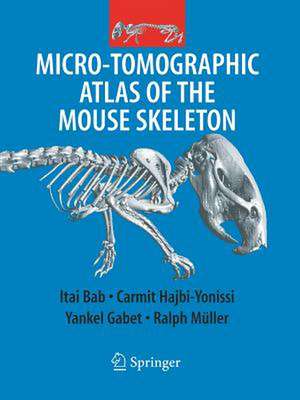
Micro-Tomographic Atlas of the Mouse Skeleton
Autor Itai A. Bab, Carmit Hajbi-Yonissi, Yankel Gabet, Ralph Mülleren Limba Engleză Paperback – 22 aug 2016
Preț: 1412.79 lei
Preț vechi: 1487.15 lei
-5% Nou
Puncte Express: 2119
Preț estimativ în valută:
270.42€ • 293.84$ • 227.30£
270.42€ • 293.84$ • 227.30£
Carte tipărită la comandă
Livrare economică 21 aprilie-05 mai
Preluare comenzi: 021 569.72.76
Specificații
ISBN-13: 9781489978929
ISBN-10: 1489978925
Pagini: 205
Ilustrații: VIII, 205 p.
Dimensiuni: 210 x 279 mm
Greutate: 0.49 kg
Ediția:Softcover reprint of the original 1st ed. 2007
Editura: Springer Us
Colecția Springer
Locul publicării:New York, NY, United States
ISBN-10: 1489978925
Pagini: 205
Ilustrații: VIII, 205 p.
Dimensiuni: 210 x 279 mm
Greutate: 0.49 kg
Ediția:Softcover reprint of the original 1st ed. 2007
Editura: Springer Us
Colecția Springer
Locul publicării:New York, NY, United States
Descriere
At the present time, the laboratory mouse has become a central tool for skeletal studies, mainly because of the extensive use of genetic manipulations in this species. Naturally, this widespread use of mice in developmental, bone, joint, tooth, and neurological research calls for detailed anatomical knowledge of the mouse skeleton as a reference for experimental design and phenotyping under a variety of experimental conditions, including genetic manipulations (e.g., transgenic and kno- out mice). Several general treatises on the normal anatomy of the mouse and rat have been published in the previous century. In the absence of adequate technologies, these books describe only the external anatomical features of the different parts of the skeleton. In general, images in these atlases are camera lucida-based line drawings rather than accurate three-dimensional images. Furthermore, so far a systematic two- and three-dimensional description of the internal anatomy of bones, as well as the three-dimensional relationship exhibited in joints, are not available.
Cuprins
Axial Skeleton.- Nose, Palate and Upper Jaw, Cranium and Tympanic Bulla.- Hyoid, Mandible, and Temporo-Mandibular Joint.- Cervical Vertebrae.- Thoracic Vertebrae.- Lumbar Vertebrae.- Sacrum.- Caudal Vertebrae.- Sternum, Sternal-Rib Joint, Ribs and Rib-Vertebral Joints.- Appendicular Skeleton.- Clavicle.- Scapula.- Humerus and Shoulder Joint.- Forearm (Ulna, Radius, and Elbow Joint).- Manus.- Pelvic Girdle.- Femur and Hip Joint.- Tibio-Fibular Complex and Knee Joint.- Hindfoot.- Murine Comparative Microanatomy.- Strain Differences.- Gender and Age Differences.
Recenzii
From the reviews:
"The format of the book is clearly defined and well organized by the authors. … serve as an important visual reference for researchers using mouse models in skeletal biology research. The Micro-Tomographic Atlas of the Mouse Skeleton is a great elementary reference to any collection and wonderfully illustrates the tremendous power of micro-CT technology in the imaging of the rodent skeleton." (Steven M. Tommasini and Christopher Price, Journal of Mammalian Evolution, Vol. 16, 2009)
"The format of the book is clearly defined and well organized by the authors. … serve as an important visual reference for researchers using mouse models in skeletal biology research. The Micro-Tomographic Atlas of the Mouse Skeleton is a great elementary reference to any collection and wonderfully illustrates the tremendous power of micro-CT technology in the imaging of the rodent skeleton." (Steven M. Tommasini and Christopher Price, Journal of Mammalian Evolution, Vol. 16, 2009)
Textul de pe ultima copertă
Micro-Tomographic Atlas of the Mouse Skeleton
Professor Itai Bab, Chief, Bone Laboratory, The Hebrew University of Jerusalem, Jerusalem, Israel
Professor Ralph Müller, Director, Center for Bioengineering Research and Education, ETH Zürich, Switzerland
Micro-Tomographic Atlas of the Mouse Skeleton serves as an essential guide containing unique systematic description of all calcified components of the mouse. This detailed atlas fulfils an emerging need for high resolution anatomical details as mice become a standard laboratory animal in skeletal research and the use of m CT technology is rapidly increasing as a key analytical tool in the study of bone.
Key Features:
- Includes over 200 high resolution, two- and three dimensional m CT images of the exterior and interiors of all bones and joints
- Offers the spatial relationship of individual bones within complex skeletal units (e.g., skull, thorax, pelvis, extremities).
- All images are accompanied by detailed explanatory text that highlights special features and newly reported structures.
- Available for the first time in the Atlas:
- Detailed information on the micro-anatomy of the murine skeleton essential for the design of experiments and interpretation of results
- Comparative analyses on m CT-based morphometric parameters at the whole bone, cortical and trabecular levels including:
- Age differences (4-40 weeks)
- Gender differences
- Differences between main mouse strains (C57Bl/6J, SJL, C3H)
Caracteristici
Detailed images accompanied by explanatory text, which describe the microstructure of the mouse skeleton.
So far a systematic two- and tri-dimensional descriptions of the internal anatomy of bones, as well as the tri-dimensional relationship exhibited in joints, is not available.
So far a systematic two- and tri-dimensional descriptions of the internal anatomy of bones, as well as the tri-dimensional relationship exhibited in joints, is not available.
