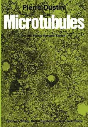
Microtubules
Cuvânt înainte de K.R. Porter Autor Pierre Dustinen Limba Engleză Paperback – 6 dec 2011
Preț: 654.12 lei
Preț vechi: 769.55 lei
-15% Nou
Puncte Express: 981
Preț estimativ în valută:
125.18€ • 130.21$ • 103.34£
125.18€ • 130.21$ • 103.34£
Carte tipărită la comandă
Livrare economică 15-29 aprilie
Preluare comenzi: 021 569.72.76
Specificații
ISBN-13: 9783642696541
ISBN-10: 3642696546
Pagini: 508
Ilustrații: XVIII, 484 p.
Dimensiuni: 170 x 244 x 27 mm
Greutate: 0.8 kg
Ediția:2nd ed. 1984. Softcover reprint of the original 2nd ed. 1984
Editura: Springer Berlin, Heidelberg
Colecția Springer
Locul publicării:Berlin, Heidelberg, Germany
ISBN-10: 3642696546
Pagini: 508
Ilustrații: XVIII, 484 p.
Dimensiuni: 170 x 244 x 27 mm
Greutate: 0.8 kg
Ediția:2nd ed. 1984. Softcover reprint of the original 2nd ed. 1984
Editura: Springer Berlin, Heidelberg
Colecția Springer
Locul publicării:Berlin, Heidelberg, Germany
Public țintă
ResearchDescriere
The decision, in 1975, to write alone a monograph on micro tubules was not without risks. While I was familiar from its start in Brussels in 1934 with the work on col chicine and other mitotic poisons, the literature on microtubules was, 8 years ago, already increasing at an impressive rate. However, this monograph, which, contrary to other works on microtubules, tried to cover the whole field of research, from the fundamentals of the tubulin molecule and the possible role of these organelles in some aspects of human pathology, to some medical applications of microtubule poisons, has been accepted as a useful tool for workers in these fields. Since 1976, (date of the last references mentioned in the monograph) until the middle of 1983, papers on microtubule research have literally been pouring in, at the rate of several hundred a year. This may justify a second edition, although the considerable difficulties in keeping the size of the book within the same limits while not forgetting to mention some important work, could not be overlooked. The need for an entirely revised and rewritten edition prompted this new venture and was possible with the help of the considerable amount of reprints kindly sent to me day after day over the years. This work would have been unthinkable if the author had not maintained the same enthusiasm for microtubule research, which has been disclosing new facts every day.
Cuprins
From the Introduction to the First Edition.- Acknowledgments.- 1 Historical Background.- 1.1 Micro tubules (MT).- 1.1.1 Definition.- 1.1.2 Early Observations.- 1.1.3 First Ultrastructural Observations.- 1.2 Colchicine: A Specific MT Poison.- 1.2.1 The Cellular Action of Colchicine.- 1.2.2 Colchicine as a Tool.- 1.2.3 Radioactive Colchicine and the Discovery of Tubulin.- 1.3 Other MT Poisons.- 1.3.1 The Catharanthus (Vinca) Alkaloids.- 1.3.2 Other Substances of Plant Origin.- 1.4 Action of Physical Agents.- 1.5 Conclusion.- References.- 2 Structure and Chemistry of Microtubules.- 2.1 Introduction.- 2.2 General Morphology of MT.- 2.3 Structure of MT.- 2.3.1 Methods of Study.- 2.3.2 The Tubulin Subunits and the MT Lattice.- 2.3.3 The Central Core.- 2.3.4 The “Exclusion” Zone and the MT “Side-Arms”.- 2.4 Biochemistry of MT and Associated Proteins.- 2.4.1 Introduction.- 2.4.2 The Tubulin Molecule.- 2.4.2.1 Amino-Acid Sequence.- 2.4.2.2 Heterogeneity.- 2.4.2.3 Post-Transcriptional Changes.- 2.4.2.4 Shape.- 2.4.2.5 Antigenicity.- 2.4.3 The Tubulin-Associated Nucleotides.- 2.4.4 The Tubulin-Associated Enzymes.- 2.4.5 The Micro tubule-Associated Proteins (HMW Proteins, MAPs, Tau).- 2.4.5.1 MAP1 and MAP2 Proteins.- 2.4.5.2 The Tau Proteins.- 2.4.5.3 Other MT-Associated Proteins.- 2.5 Assembly and Disassembly of MT in Vitro.- 2.5.1 General Conditions Required for Assembly in Vitro. Significance of “Rings”.- 2.5.2 Role of MT-Associated Proteins.- 2.5.3 Assembly Without Associated Proteins.- 2.5.4 Role of Guanine Nucleotides in Assembly.- 2.5.5 Guanine Nucleotides, “Treadmilling” and “Polarity“.- 2.5.6 Thermodynamics of Assembly.- 2.5.7 Polymorphism of Assembly.- 2.5.8 Site-Initiated Assembly in Vitro.- 2.6 Summary.- References.- 3 General Physiology of Tubulins and Microtubules.- 3.1 Introduction.- 3.2 Tubulin Synthesis and Its Regulation.- 3.2.1 In Oogenesis and Early Stages of Development.- 3.2.2 In Adult Cells.- 3.2.3 In Relation with Ciliary Growth.- 3.2.4 Role of Hormones.- 3.3 Assembly of MT in Vivo.- 3.3.1 Role of Intracellular MTOC.- 3.3.2 Role of Calcium and Calmodulin.- 3.3.3 Other Regulatory Factors of Assembly.- 3.3.4 Thermodynamic Aspects.- 3.3.5 Differential Sensitivity of MT.- 3.3.6 Abnormal Assemblies in Vivo: Macro- and Megatubules.- 3.3.7 How do MT Assemble in the Cell?.- 3.4 Relations of MT with Other Cell Structures and Organelles.- 3.4.1 Cell Membranes.- 3.4.2 Nuclear Envelope.- 3.4.3 Mitochondria.- 3.4.4 Golgi Apparatus.- 3.4.5 Lysosomes.- 3.4.6 Ribosomes.- 3.4.7 Other Cell Components.- 3.5 Relations with Viruses and Endocellular Parasites.- 3.6 Extracellular MT.- References.- 4 Complex Microtubule Assemblies: Axonemes, Centrioles, Basal Bodies, Cilia, and Flagella.- 4.1 Introduction.- 4.2 Axonemes.- 4.3 Centrioles.- 4.3.1 Definition and Number.- 4.3.2 Ultrastructure.- 4.3.3 Biochemistry.- 4.3.4 Replication and Growth.- 4.3.5 Centriole Neoformation.- 4.3.6 Atypical Centrioles.- 4.3.7 Stability of the Centriolar MT.- 4.4 Cilia and Flagella. Introduction.- 4.4.1 Structure and Chemistry.- 4.4.2 The Nine Peripheral Doublets.- 4.4.3 The Central Pair MT.- 4.4.4 The Dynein Arms.- 4.4.5 Other Ciliary Structures.- 4.5 Basal Bodies.- 4.5.1 Structure.- 4.5.2 Formation.- 4.6 Regeneration of Cilia and Ciliogenesis.- 4.7 Atypical and Pathological Cilia.- References.- 5 Microtubule Poisons.- 5.1 Introduction.- 5.2 Colchicine and Colchicine Derivatives.- 5.2.1 Chemical Structure.- 5.2.2 The Tubulin-Colchicine Bond.- 5.2.3 Action of Colchicine on MT.- 5.2.4 Changes of IMF.- 5.2.5 Colchicine-Resistant Tubulins.- 5.2.6 Action on Nucleic Acid Metabolism.- 5.2.7 Colchicine Antagonists.- 5.2.8 Colchicine Pharmacology.- 5.3 The Catharanthus (Vinca) Alkaloids.- 5.3.1 Chemical Structure.- 5.3.2 Action on Tubulin and MT.- 5.3.3 The Vinca Crystals.- 5.3.4 Other Actions of Vinca Alkaloids.- 5.4 Podophyllotoxin and Related Molecules.- 5.5 Sulfhydryl Reagents.- 5.6 The Benzimidazole Derivatives.- 5.7 Griseofulvin.- 5.8 Anesthetic Drugs.- 5.9 Other MT Poisons.- 5.10 Taxol: An Agent That Favorizes MT Assembly.- 5.11 Action of Physical Agents and Heavy Water.- References.- 6 Cell Shape.- 6.1 Introduction.- 6.2 Disk-Shaped Blood Cells.- 6.2.1 Red Blood Cells.- 6.2.2 Blood Platelets and Thrombocytes.- 6.3 Nuclear and Cytoplasmic Shaping in Spermatogenesis.- 6.4 Other Cytoskeletal Functions of MT in Metazoa.- 6.4.1 Introduction.- 6.4.2 Epithelial Cells.- 6.4.3 Connective Tissue Cells.- 6.4.4 Schwann Cells and Glia.- 6.5 Egg Differentiation and Embryonic Growth.- 6.6 Cell Shape in Plants.- 6.6.1 MT and Cellulose Microfilaments.- 6.6.2 Are There MTOC in Plant Cells?.- 6.6.3 The Preprophase Band in Plant Mitoses.- 6.7 Cell Shape in Protozoa.- 6.8 MT with Mechanical Functions.- References.- 7 Cell Movement.- 7.1 Introduction.- 7.2 Intracellular Displacements and Motion.- 7.2.1 Saltatory Motion.- 7.2.2 Axoneme-Associated Movements.- 7.2.3 Movements Associated with Feeding in Simple Organisms.- 7.2.4 Other Intracytoplasmic Movements Related to MT.- 7.3 The Movement of Pigment Granules.- 7.4 Movements of Cell Membranes and “Capping”.- 7.4.1 Capping in Lymphocytes.- 7.4.2 In Polymorphonuclear Leukocytes.- 7.4.3 Capping in Other Cells.- 7.5 Cell Motility and Locomotion.- 7.5.1 Polymorphonuclear Leukocytes.- 7.5.2 Mononuclears and Macrophages.- 7.5.3 Lymphocytes.- 7.5.4 Other Cells.- 7.6 Ciliary Movements.- References.- 8 Secretion.- 8.1 Introduction.- 8.2 Endocrine Secretions.- 8.2.1 The Langerhans Islets.- 8.2.2 The Thyroid.- 8.2.3 The Parathyroid.- 8.2.4 Pituitary, Anterior Lobe.- 8.2.5 The Adrenals.- 8.3 Exocrine Secretions.- 8.3.1 Pancreas.- 8.3.2 Salivary and Lacrymal Glands.- 8.3.3 Mammary Gland and Milk Secretions.- 8.3.4 Gastric and Intestinal Glands.- 8.3.5 Hepatic Cells and Liver Secretions.- 8.4 Leukocytes and Related Cells.- 8.4.1 Polymorphonuclear Leukocytes (PMN).- 8.4.2 Basophils and Mastcells.- 8.4.3 Monocytes and Macrophages.- 8.4.4 Plasmatocytes and the Secretion of Immunoglobulins.- 8.5 Other Cell Activities Related to Secretion.- 8.5.1 Fibroblasts, Osteoblasts, Chondrocytes, Ameloblasts, and Collagen Secretion.- 8.5.2 Other Cells.- 8.6 Conclusions.- References.- 9 Neurotubules and Neuroplasmic Transport.- 9.1 Introduction.- 9.2 General Properties of Nerve MT.- 9.3 Relations of MT with Synaptic Vesicles.- 9.4 Microtubules and Nerve Cells Shape.- 9.5 Experimental Changes of Neuronal MT.- 9.6 Neuroplasmic Transport and MT.- 9.6.1 Introduction.- 9.6.2 Technical Approaches.- 9.6.3 Orthograde Flow.- 9.6.4 Retrograde Flow.- 9.6.5 The Transport of Neurosecretory Granules.- 9.6.6 Fate of the Transported Metabolites and of MT.- 9.7 Role of MT in Axoplasmic Transport.- 9.7.1 Colchicine.- 9.7.2 Other MT Poisons.- 9.7.3 Action of Cold and Heavy Water.- 9.8 Theories of Neuroplasmic Transport.- 9.9 MT and Sensory Cells.- References.- 10 Microtubules and Mitosis.- 10.1 Introduction.- 10.2 Some Aspects of the Possible Evolution of Mitosis.- 10.3 Some Types of Mitosis.- 10.3.1 Intranuclear MT.- 10.3.2 Partially Intranuclear MT.- 10.3.3 Extranuclear Spindles Attached to Nuclear Membrane and Chromosomes.- 10.3.4 Mitoses in Higher Plants and Animals.- 10.4 Methods of Study.- 10.5 MT and Mitotic Movements.- 10.5.1 Prophase.- 10.5.2 From Prophase to Metaphase.- 10.5.3 Anaphase.- 10.5.4 Telophase.- 10.6 Other Proteins Associated with Mitotic MT.- 10.6.1 Associated Proteins: MAPs, Tau Factor.- 10.6.2 Microfilaments: Actin, Myosin, Dynein.- 10.6.3 Calcium and Calmodulin.- 10.7 The Action of MT Poisons on Mitosis.- 10.7.1 General Aspects.- 10.7.2 Colchicine.- 10.7.3 Other Chemical MT Poisons.- 10.7.4 Taxol.- 10.7.5 Deuterium Oxide.- 10.8 The Action of Physical Factors on Mitosis.- 10.8.1 Cold.- 10.8.2 Heat.- 10.8.3 The Local Action of Ultraviolet Microbeams.- 10.8.4 High Hydrostatic Pressure.- 10.9 Microtubules and the Mechanisms of Mitosis.- 10.9.1 Introduction.- 10.9.2 Mitotic Movements in Isolated MA.- 10.9.3 The Role of Assembly-Disassembly.- 10.9.4 The Sliding-Filament Concept.- 10.9.5 “Zipping”.- 10.9.6 The Possible Role of Treadmilling.- 10.10 Conclusions.- References.- 11 MT and MT Poisons in Pathology and Medicine.- 11.1 Introduction.- 11.2 Pathology of MT Structures.- 11.2.1 Abnormal Cilia.- 11.2.2 Other Pathological Conditions.- 11.2.3 MT in Transformed, Neoplastic Cells.- 11.3 Therapeutic Uses of MT Poisons.- 11.3.1 Introduction.- 11.3.2 Colchicine.- 11.3.3 Colchicine Poisoning in Man.- 11.4 The Toxicity of the Vinca (Catharanthus) Alkaloids.- 11.4.1 Blood Platelets.- 11.4.2 Neurological Disturbances.- 11.5 Toxicity of MT Poisons in Cancer Chemotherapy 433 References.- 12 Post-Script and Outlook.- 12.1 Unity and Diversity.- 12.2 Microtubule-Associated Proteins (MAPs).- 12.3 Assembly and Disassembly.- 12.4 Microtubule Poisons.- 12.5 Non-Microtubular Tubulin.- 12.6 Micro tubules and Movement.- 12.7 Micro tubules and Cell Shape.- 12.8 Micro tubules and Evolution.- References.- Addenda Some of the Most Interesting Papers Published in 1983 and Early in 1984.- Recent Books and Reviews on Microtubules and Related Subjects.


