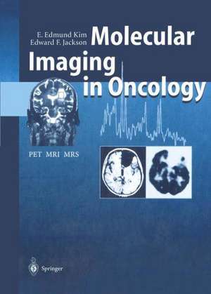
Molecular Imaging in Oncology: PET, MRI, and MRS
Autor E. Edmund Kim Contribuţii de J. Aoki Autor Edward F. Jackson Contribuţii de H. Baghaei, S. Ilgan, T. Inoue, H. Li, J. Uribe, F.C.L. Wong, W.-H. Wong, D.J. Yangen Limba Engleză Paperback – 30 sep 2011
Preț: 684.06 lei
Preț vechi: 720.07 lei
-5% Nou
Puncte Express: 1026
Preț estimativ în valută:
130.90€ • 137.01$ • 108.95£
130.90€ • 137.01$ • 108.95£
Carte tipărită la comandă
Livrare economică 26 martie-01 aprilie
Preluare comenzi: 021 569.72.76
Specificații
ISBN-13: 9783642641633
ISBN-10: 3642641636
Pagini: 308
Ilustrații: XIII, 290 p. 5 illus. in color.
Dimensiuni: 193 x 270 x 16 mm
Greutate: 0.67 kg
Ediția:Softcover reprint of the original 1st ed. 1999
Editura: Springer Berlin, Heidelberg
Colecția Springer
Locul publicării:Berlin, Heidelberg, Germany
ISBN-10: 3642641636
Pagini: 308
Ilustrații: XIII, 290 p. 5 illus. in color.
Dimensiuni: 193 x 270 x 16 mm
Greutate: 0.67 kg
Ediția:Softcover reprint of the original 1st ed. 1999
Editura: Springer Berlin, Heidelberg
Colecția Springer
Locul publicării:Berlin, Heidelberg, Germany
Public țintă
ResearchCuprins
Principles and Technology.- 1 Principles of Cancer Biology, Biochemistry, Immunology and Pathology.- 2 Imaging Strategies and Perspectives in Oncology.- 3 Magnetic Resonance Imaging: Physical Principles to Advanced Applications.- 4 Magnetic Resonance Spectroscopy: Physical Principles and Applications.- 5 Principles and Instrumentation of Position Emission Tomography.- 6 Radiopharmaceuticals for Tumor Imaging and Magnetic Resonance Imaging Contrast Agents.- 7 Receptor Imaging.- 8 Practical Magnetic Resonance Imaging and Positron Emission Tomography Techniques and Their Artifacts.- Clinical Applications of MRI, MRS and PET.- 9 Lung Cancers.- 10 Breast Cancer.- 11 Gastrointestinal Carcinomas.- 12 Urologic Cancers.- 13 Gynecologic Cancers.- 14 Brain Tumors.- 15 Head and Neck Tumors.- 16 Musculoskeletal Tumors.- 17 Melanoma, Lymphoma and Myeloma.
Recenzii
From the reviews:
“This handbook focuses on the growing impact of molecular imaging in oncology and addresses topics ranging from basic research to clinical applications in the era of evidence-based medicine. … I highly recommend this book to nuclear physicians, radiologists, oncologists, chemists, physicists, mathematicians, and computer scientists.” (E. Edmund Kim, The Journal of Nuclear Medicine, Vol. 54 (6), June, 2013)
“This handbook focuses on the growing impact of molecular imaging in oncology and addresses topics ranging from basic research to clinical applications in the era of evidence-based medicine. … I highly recommend this book to nuclear physicians, radiologists, oncologists, chemists, physicists, mathematicians, and computer scientists.” (E. Edmund Kim, The Journal of Nuclear Medicine, Vol. 54 (6), June, 2013)
Textul de pe ultima copertă
Advanced imaging techniques often make it possible to diagnose localized abnormalities prior to irreversible damage. PET permits visualization of tumor metabolic or physiologic characteristics. MRI shows morphologic abnormalities and allows assessment of the functional status of tissue, including the ability to indirectly map areas of task-induced neuronal activation and to map blood volume and flow. MRS provides noninvasive biochemical information from tumors and surrounding normal tissue. By combining PET, MRI and MRS information we should be able to differentiate tumors from non-tumor lesions, characterize types or grades of tumors, monitor tumor regression, recurrence or response to therapy, and also image the location of gene delivery. This book reports updated techniques, instrumentation and clinical application of PET, MRI and MRS in cancer management.
Caracteristici
Includes supplementary material: sn.pub/extras
