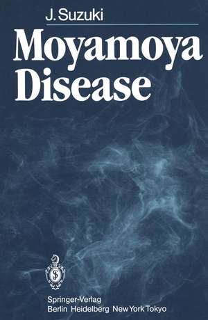
Moyamoya Disease
Autor Jiro Suzukien Limba Engleză Paperback – apr 1986
Preț: 712.25 lei
Preț vechi: 749.73 lei
-5% Nou
Puncte Express: 1068
Preț estimativ în valută:
136.33€ • 148.14$ • 114.59£
136.33€ • 148.14$ • 114.59£
Carte tipărită la comandă
Livrare economică 21 aprilie-05 mai
Preluare comenzi: 021 569.72.76
Specificații
ISBN-13: 9783540157786
ISBN-10: 3540157786
Pagini: 204
Ilustrații: XII, 190 p.
Dimensiuni: 170 x 244 x 11 mm
Greutate: 0.33 kg
Editura: Springer Berlin, Heidelberg
Colecția Springer
Locul publicării:Berlin, Heidelberg, Germany
ISBN-10: 3540157786
Pagini: 204
Ilustrații: XII, 190 p.
Dimensiuni: 170 x 244 x 11 mm
Greutate: 0.33 kg
Editura: Springer Berlin, Heidelberg
Colecția Springer
Locul publicării:Berlin, Heidelberg, Germany
Public țintă
ResearchCuprins
1 History and Definition.- 2 Epidemiology and Symptomatology.- 2.1 Introduction.- 2.2 Incidence.- 2.3 Incidence by Sex.- 2.4 Age at Onset.- 2.5 Age on Admission.- 2.6 Initial Symptoms and Symptoms over the Clinical Course.- 2.7 Laboratory Findings on Admission.- 2.8 Family History.- 2.9 Anamnesis.- 2.10 Conclusion.- 3 Cerebral Angiography.- 3.1 Introduction.- 3.2 Basal Moyamoya.- 3.3 Nature of Basal Moyamoya.- 3.4 Ethmoidal Moyamoya.- 3.5 Vault Moyamoya.- 3.6 Cerebral Angiograms Taken After Hyperventilation.- 3.7 Summary.- 4 Mechanisms of Symptomatic Occurrence.- 4.1 Introduction.- 4.2 Mechanism of TIA in Children.- 4.3 Mechanism of Onset of Symptoms Due to Intracranial Hemorrhage in Adults.- 4.4 Summary.- 5 Electroencephalography.- 5.1 Introduction.- 5.2 Electrical Activity of the Brain in Juvenile Moyamoya Disease.- 5.3 Electroencephalography in Adult Cases of Moyamoya Disease.- 5.4 Evoked Potentials.- 5.5 Summary.- 6 Computerized Tomography.- 6.1 Introduction.- 6.2 Unenhanced Scan.- 6.3 Enhanced Scan.- 6.4 Stage of Moyamoya Disease and Computerized Tomography.- 6.5 Conclusion.- 7 Positron Emission Computerized Tomography.- 7.1 Introduction.- 7.2 Cases.- 8 Cerebral Hemodynamics.- 8.1 Introduction.- 8.2 Average Cerebral Blood Flow and Hemispherical Blood Flow.- 8.3 Vascular Response in Moyamoya Disease.- 8.4 Conclusion.- 9 Treatment.- 9.1 Introduction.- 9.2 Therapy of Infarction Attacks.- 9.3 Superficial Temporal Artery-Cortical Branch of the Middle Cerebral Artery Anastomosis (STA-MCA Anastomosis) and Encephalomyo-synangiosis.- 9.4 Transplantation of the Omentum.- 9.5 Encephaloduroarteriosynangiosis.- 9.6 Therapy for and Prevention of Intracranial Hemorrhage.- 9.7 Anesthesia During Surgery for Moyamoya Disease.- 9.8 Conclusion.- 10 Pathology.- 10.1 Introduction.- 10.2Autopsy Materials.- 10.3 Pathology of Autopsied Brain Tissue.- 10.4 Investigations Based upon Whole Body Autopsy.- 10.5 Conclusion.- 11 Etiology.- 11.1 Introduction.- 11.2 Background to Our Experimental Research.- 11.3 Experimental Methods and Results.- 11.4 Conclusion.- 12 Quasi-Moyamoya Diseases.- 12.1 Introduction.- 12.2 Unilateral Quasi-Moyamoya Disease.- 12.3 Quasi-Moyamoya Disease in Which Bilateral or Unilateral Stenosis or Occlusion of the Proximal Portion of the MCA Is Seen, Together with Moyamoya Vessels in the Immediate Vicinity.- 12.4 Quasi-Moyamoya Disease with Vascular Malformations.- 12.5 Quasi-Moyamoya Disease Due to Known Causes.- 12.6 Quasi-Moyamoya Disease Due to Other Causes.- 12.7 Conclusion.- References.
