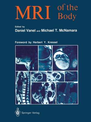
MRI of the Body
Editat de Daniel Vanel Prefață de Herbert Y. Kressel Traducere de Susanne Assenat Editat de Michael T. McNamaraen Limba Engleză Paperback – 17 aug 2012
Preț: 736.03 lei
Preț vechi: 774.76 lei
-5% Nou
Puncte Express: 1104
Preț estimativ în valută:
140.86€ • 146.51$ • 116.29£
140.86€ • 146.51$ • 116.29£
Carte tipărită la comandă
Livrare economică 12-26 aprilie
Preluare comenzi: 021 569.72.76
Specificații
ISBN-13: 9783642875588
ISBN-10: 3642875580
Pagini: 412
Ilustrații: XXIII, 387 p. 1000 illus., 9 illus. in color.
Dimensiuni: 210 x 279 x 22 mm
Greutate: 0.92 kg
Ediția:Softcover reprint of the original 1st ed. 1989
Editura: Springer Berlin, Heidelberg
Colecția Springer
Locul publicării:Berlin, Heidelberg, Germany
ISBN-10: 3642875580
Pagini: 412
Ilustrații: XXIII, 387 p. 1000 illus., 9 illus. in color.
Dimensiuni: 210 x 279 x 22 mm
Greutate: 0.92 kg
Ediția:Softcover reprint of the original 1st ed. 1989
Editura: Springer Berlin, Heidelberg
Colecția Springer
Locul publicării:Berlin, Heidelberg, Germany
Public țintă
ResearchCuprins
Physical basis.- Physical basis of nuclear magnetic resonance.- Signal parameters.- Formation of an image.- Artifacts.- System-specific artifacts.- Patient-specific artifacts.- Quality control.- Definition of QC parameters.- Test substances and test objects.- NMR spectroscopy from experimental to clinical spectroscopy.- Principle of NMR spectroscopy.- Most significant results of spectrocopy in man.- Clinical application.- Conclusion.- Contrast media.- Theoretic basis.- Paramagnetic ions.- Other contrast media.- Experimental models.- Clinical applications.- Conclusion.- Head and neck.- Facial structures — nasopharynx and parapharyngeal spaces.- Superficial soft tissue (excluding the orbits) : parotid gand and temporomandibular joint.- The parotid gland.- Buccal cavity and the oropharynx.- Cervical region.- Conclusion.- Thorax.- Exploration techniques.- Normal anatomy.- Pathological findings.- Conclusion.- Heart.- General points.- Study of the heart.- Clinical applications.- Breast.- Imaging technique.- MR image of the normal breast.- Results.- Conclusion.- Liver, biliary tract, portal system, spleen.- Technique.- Normal anatomy.- Clinical findings.- Conclusion.- Pancreas.- Application of MR imaging techniques to the pancreas.- Normal pancreas.- Acute and chronic pancreatitis.- Liquid collections and pseudocysts.- Pancreatic hemorrhage.- Tumors of the pancreas.- Metabolic diseases.- Vascular abnormalities associated with hepatic diseases.- Present situation, prospects, comparison with CT.- Tissue characterizarion.- Gastrointestinal tract.- MR examination technique for the GI tract.- Normal anatomy of the rectum.- Abnormalities of the GI tract.- Advantages and disadvantages of MR — Future prospects.- The kidneys and perirenal space.- Technique.- Anatomy.- Masslesions.- Loss of corticomedullary differentiation.- Perirenal lesions.- Paramagnetic substances.- Adrenal glands.- MRI procedure.- Normal anatomy.- Secretory tumors of the adrenals.- Non-secretory tumors.- Other lesions.- Contribution of spectroscopy imaging to adrenal investigation.- Conclusion.- Large retroperitoneal blood vessels.- Exploration technique.- Normal findings.- Pathological findings.- Conclusion.- Retroperitoneal adenopathy.- Technique.- Findings.- Conclusion.- Gynecology.- Examination technique.- Normal anatomy.- Benign pathology.- Malignant pathology.- Postoperative pathology and therapeutic follow-up.- Conclusion.- Male pelvis.- Technique.- Normal anatomy.- Pathology.- Conclusion.- Pathology of the scrotum.- Examination technique.- Normal appearance.- Pathology of the scrotum.- Conclusion.- Joints.- General technical points.- Normal images.- General findings.- Pathological conditions.- Spine.- Technical considerations.- Normal images.- Degenerative pathology.- Infections of the disk and of the vertebra.- Spondylolysis and spondylolisthesis.- Trauma.- Inflammatory pathology.- Post-treatment appearance of the spine.- Spinal tumors.- Conclusion.- Primary musculoskeletal tumors.- Advantages and limitations of MRI.- Technique.- Contribution of MRI to diagnosis.- Assessment of tumor extension.- Treatment efficacy.- Post-treatment checkup.- Practical examples.- Conclusion.- Bone Marrow: MRI of diffuse and multifocal bone marrow malignancy.- Technique.- Normal bone marrow.- Pathological bone marrow.- Conclusion.- Role of MR in non-oncologic pediatric imaging.- Technique.- Indications.- Discussion.- Conclusion.- MRI in pediatric oncology.- Material and techniques.- Tumoral pathology.- Conclusion.- Obstetrical MRI.- The mother.- The fetus.- Application of MRIto radiation therapy.- Contribution of MRI to radiation therapy planning.- Geometric distortion.


