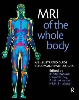
MRI of the Whole Body: An Illustrated Guide for Common Pathologies
Autor Nikhil Bhuskute, Edward Hoey, Amit Lakkaraju, Kshitij Mankaden Limba Engleză Paperback – 30 sep 2011
MRI of the Whole Body sets out to educate trainee and experienced radiologists, radiographers and clinicians regarding key sequences for optimal imaging of common pathologies, with simple explanations on the choice of a particular MR sequence. The authors present typical and representative examples with relevant clinical and imaging features to assist a better understanding of these commonly encountered conditions. Every unit begins with a quick anatomy review, and each case is described in a standardised format with a clinical background, key sequences, imaging features, and practical hints as to close differentials and ways to distinguish between them. A text of this nature is essential for all MR practitioners whatever their background: medical, technical or scientific.
Key features:
- First of its kind as no other book covers all body systems in one volume with demonstration of all key imaging sequences in the commonly diagnosed pathologies
- Up-to-date sequences described with reasons for choosing a particular sequence for a particular case
- Simplified relevant MR anatomy preceding each unit
- Clear high resolution images with appropriate legends
- Practical hints and tips section included for each pathology - close differentials and what to do next
Preț: 364.71 lei
Preț vechi: 465.55 lei
-22% Nou
Puncte Express: 547
Preț estimativ în valută:
69.79€ • 73.05$ • 58.09£
69.79€ • 73.05$ • 58.09£
Carte tipărită la comandă
Livrare economică 31 martie-14 aprilie
Preluare comenzi: 021 569.72.76
Specificații
ISBN-13: 9781853157769
ISBN-10: 1853157767
Pagini: 276
Ilustrații: 10; 600 photos
Dimensiuni: 219 x 276 x 16 mm
Greutate: 0.78 kg
Ediția:ILL
Editura: CRC Press
Colecția CRC Press
ISBN-10: 1853157767
Pagini: 276
Ilustrații: 10; 600 photos
Dimensiuni: 219 x 276 x 16 mm
Greutate: 0.78 kg
Ediția:ILL
Editura: CRC Press
Colecția CRC Press
Public țintă
Professional ReferenceCuprins
Unit I MRI of the Musculoskeletal System: Musculoskeletal Imaging. The Spine. The Knee. The Hip. The Shoulder. The Wrist. The Foot and Ankle. Malignancy and Infection. Unit II Neuro-MRI: Sequences and Protocols in Neuri-MRI. Neuroanatomy. Vascular Pathologies. Neoplasms. Developmental Conditions. Vasculitis/Demyelination. Infections. Congenital Conditions. Unit III: Anatomy. Abdominal. Urology and Gynaecology. Unit IV: Key Sequences. Cardiothoracic Pathologies.
Notă biografică
Kshitij Mankad MRCP FRCR Neuroradiology Fellow, Barts and the London NHS Trust, London, UK; Edward TD Hoey BAO MRCP FRCR Consultant Cardiothoracic Radiologist, Heart of England Foundation Trust; Honorary Senior Clinical Lecturer, University of Birmingham Medical School, Birmingham, UK; Amit Lakkaraju FRCR PG MED ED Consultant Musculoskeletal Radiologist, Goulburn Valley Base Hospital, Shepparton, Australia; Nikhil Bhuskute MS FRCS FRCR Consultant Radiologist, Calderdale and Huddersfield NHS Foundation Trust, UK
Descriere
Designed for trainee and experienced radiologists, radiographers and clinicians, this book explains key sequences for optimal imaging of common pathologies, with simple explanations on the choice of a particular MR sequence. The authors present representative examples to facilitate a better understanding of these commonly encountered conditions. Each case is described in a standardized format with clinical background, key sequences, imaging features, and practical hints as to close differentials and ways to distinguish between them. This text is essential for all MR practitioners whether their background is medical, technical, or scientific.
