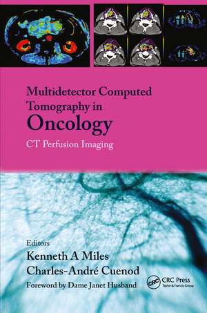
Multi-Detector Computed Tomography in Oncology: CT Perfusion Imaging
Editat de Kenneth Miles, C. Charnsangavej, C. Cuenoden Limba Engleză Hardback – 15 sep 2007
Short Contents
Preț: 1065.24 lei
Preț vechi: 1294.29 lei
-18% Nou
Puncte Express: 1598
Preț estimativ în valută:
203.84€ • 210.29$ • 170.10£
203.84€ • 210.29$ • 170.10£
Carte tipărită la comandă
Livrare economică 27 martie-10 aprilie
Preluare comenzi: 021 569.72.76
Specificații
ISBN-13: 9781842143094
ISBN-10: 1842143093
Pagini: 258
Ilustrații: 34 b/w images, 39 color images, 19 color tables, 34 halftones and 39 color halftones
Dimensiuni: 156 x 234 x 21 mm
Greutate: 0.75 kg
Ediția:1
Editura: CRC Press
Colecția CRC Press
ISBN-10: 1842143093
Pagini: 258
Ilustrații: 34 b/w images, 39 color images, 19 color tables, 34 halftones and 39 color halftones
Dimensiuni: 156 x 234 x 21 mm
Greutate: 0.75 kg
Ediția:1
Editura: CRC Press
Colecția CRC Press
Public țintă
Professional ReferenceCuprins
1. Acquisition protocols - K.A. Miles
2. Image-processing software - M.R. Griffiths [Australia]
3. Pathophysiology of tumour vasculature - C. Charnsangavej
4. Head & neck cancer - R. Hermans [Belgium]
5. Lung cancer - U. Tateishi [Japan]
6. Liver metastases - C. Cuenod & K.A. Miles
7. Prostate cancer - Swithin Song [Australia]
8. Other tumours: lymphoma, kidney, pancreas - K.A. Miles
9. Beyond RECIST - Perfusion in CT in cancer trials - C. Charnsangavej & K.A. Miles
10. Perfusion CT for CT/PET systems - K.A. Miles
Notă biografică
Kenneth Miles, C. Charnsangavej, C. Cuenod
Descriere
This new text-atlas focuses on anatomy and procedural strategy for perfusion CT imaging in the diagnosis and management of cancer. It will use a combination of pictures and schematic diagrams that show how this new modality can be used to assess anatomy and guide therapeutic interventions. It begins with an introductory section discussing the state of the art and background support (including software) in the use of the technique; there then follows a sequence of chapters that review applications for each of the main body systems and anatomic regions. The book concludes with a section on the uses of perfusion CT in monitoring clinical trials, and also reviews new applications for combined modalities such as CT/PET.
