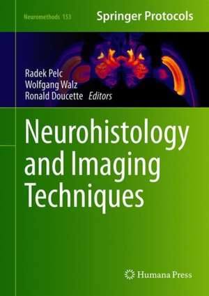
Neurohistology and Imaging Techniques: Neuromethods, cartea 153
Editat de Radek Pelc, Wolfgang Walz, J. Ronald Doucetteen Limba Engleză Hardback – 20 noi 2020
This volume explores major light microscopic imaging modalities that can be used to view nervous tissue, and discusses the steps needed to use each of them, and ways to interpret the data. The chapters in this book cover topics such as atlasing of insect brain; neuroanatomical tracing through fluorochrome expression; fluorescent probes for amyloids; or optical clearing for ultramicroscopy of GFP-expressing tissues. In the Neuromethods series style, chapters include the kind of detail and key advice from the specialists needed to get successful results in your laboratory.
Authoritative and cutting-edge, Neurohistology and Imaging Techniques is a valuable resource for both expert and novice users of major light microscopic imaging techniques, and those interested in exploring alternate imaging tools.
Din seria Neuromethods
- 5%
 Preț: 347.57 lei
 - 15%
 Preț: 659.53 lei
 - 15%
 Preț: 665.08 lei
 - 18%
 Preț: 986.63 lei
 - 24%
 Preț: 852.89 lei
 - 18%
 Preț: 953.03 lei
 - 18%
 Preț: 955.25 lei
 - 20%
 Preț: 1129.36 lei
 - 20%
 Preț: 1252.04 lei
 - 18%
 Preț: 1291.45 lei
 - 15%
 Preț: 652.31 lei
 - 18%
 Preț: 955.70 lei
 - 23%
 Preț: 705.39 lei
 - 18%
 Preț: 973.38 lei
 - 18%
 Preț: 964.86 lei
 - 18%
 Preț: 968.03 lei
 - 15%
 Preț: 662.95 lei
 - 15%
 Preț: 646.43 lei
 - 15%
 Preț: 649.71 lei
 -
 Preț: 395.29 lei
 - 19%
 Preț: 580.67 lei
 - 19%
 Preț: 584.12 lei
 - 19%
 Preț: 566.41 lei
 - 15%
 Preț: 652.17 lei
 - 15%
 Preț: 655.13 lei
 - 18%
 Preț: 959.36 lei
 - 15%
 Preț: 652.49 lei
 - 15%
 Preț: 649.54 lei
 - 15%
 Preț: 649.87 lei
 - 15%
 Preț: 650.19 lei
 - 15%
 Preț: 648.42 lei
 - 18%
 Preț: 1039.22 lei
 - 18%
 Preț: 963.15 lei
Preț: 966.27 lei
Preț vechi: 1178.38 lei
-18% Nou
Puncte Express: 1449
Preț estimativ în valută:
184.89€ • 193.56$ • 152.99£
184.89€ • 193.56$ • 152.99£
Carte tipărită la comandă
Livrare economică 05-19 aprilie
Preluare comenzi: 021 569.72.76
Specificații
ISBN-13: 9781071604267
ISBN-10: 1071604260
Pagini: 472
Ilustrații: XIV, 472 p. 159 illus., 105 illus. in color.
Dimensiuni: 178 x 254 mm
Greutate: 1.04 kg
Ediția:1st ed. 2020
Editura: Springer Us
Colecția Humana
Seria Neuromethods
Locul publicării:New York, NY, United States
ISBN-10: 1071604260
Pagini: 472
Ilustrații: XIV, 472 p. 159 illus., 105 illus. in color.
Dimensiuni: 178 x 254 mm
Greutate: 1.04 kg
Ediția:1st ed. 2020
Editura: Springer Us
Colecția Humana
Seria Neuromethods
Locul publicării:New York, NY, United States
Cuprins
Neurohistology with a Touch of History: Technology-Driven Research.- Fixation Protocols for Neurohistology: Neurons to Genes.- Three-Dimensional Atlases of Insect Brains.- Neuroanatomical Tracing Based on Selective Fluorochrome Expression in Transgenic Animals.- Optical Imaging Probes for Amyloid Diseases in Brain.- Chemical Clearing of GFP-Expressing Neural Tissues.- The Properties of Light Governing Biological Microscopy.- Beyond Brightfield: “Forgotten” Microscopic Modalities.- Stereomicroscopy in Neuroanatomy.- Conventional, Apodized, and Relief Phase-Contrast Microscopy.- Ultramicroscopy of Nerve Fibers and Neurons: Fine-Tuning the Light Sheets.- Imaging and Electrophysiology of Individual Neurites Functionally Isolated in Microchannels.- Consumer Versus Dedicated Digital Cameras in Photomicrography.- Digital Micrographs in Pathology.
Textul de pe ultima copertă
This volume explores major light microscopic imaging modalities that can be used to view nervous tissue, and discusses the steps needed to use each of them, and ways to interpret the data. The chapters in this book cover topics such as atlasing of insect brain; neuroanatomical tracing through fluorochrome expression; fluorescent probes for amyloids; or optical clearing for ultramicroscopy of GFP-expressing tissues. In the Neuromethods series style, chapters include the kind of detail and key advice from the specialists needed to get successful results in your laboratory. Authoritative and cutting-edge, Neurohistology and Imaging Techniques is a valuable resource for both expert and novice users of major light microscopic imaging techniques, and those interested in exploring alternate imaging tools.
Caracteristici
Includes cutting-edge methods and protocols Provides step-by-step detail essential for reproducible results Contains key notes and implementation advice from the experts
