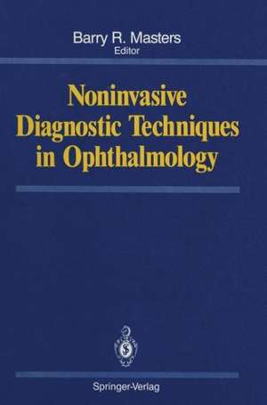
Noninvasive Diagnostic Techniques in Ophthalmology
Editat de Barry R. Masters Cuvânt înainte de David Mauriceen Limba Engleză Paperback – 19 ian 2012
Preț: 398.91 lei
Preț vechi: 419.91 lei
-5% Nou
Puncte Express: 598
Preț estimativ în valută:
76.33€ • 80.12$ • 63.36£
76.33€ • 80.12$ • 63.36£
Carte tipărită la comandă
Livrare economică 10-24 aprilie
Preluare comenzi: 021 569.72.76
Specificații
ISBN-13: 9781461388982
ISBN-10: 1461388988
Pagini: 684
Ilustrații: XXXII, 649 p.
Dimensiuni: 178 x 254 x 36 mm
Greutate: 1.17 kg
Ediția:Softcover reprint of the original 1st ed. 1990
Editura: Springer
Colecția Springer
Locul publicării:New York, NY, United States
ISBN-10: 1461388988
Pagini: 684
Ilustrații: XXXII, 649 p.
Dimensiuni: 178 x 254 x 36 mm
Greutate: 1.17 kg
Ediția:Softcover reprint of the original 1st ed. 1990
Editura: Springer
Colecția Springer
Locul publicării:New York, NY, United States
Public țintă
ResearchCuprins
1 Ophthalmic Image Processing.- 2 Magnetic Resonance Imaging of the Eye and Orbit.- 3 Magnetic Resonance Imaging in Ophthalmology.- 4 Diagnostic Ocular Ultrasonography.- 5 Corneal Topography.- Appendix: Considerations in Corneal Surface Reconstruction from Keratoscope Images.- 6 Holographic Contour Analysis of the Cornea.- 7 Wide-Field Specular Microscopy.- 8 Fourier Transform Method for Statistical Evaluation of Corneal Endothelial Morphology.- 9 Color Specular Microscopy.- 10 Confocal Microscopy of the Eye.- 11 Confocal Microscopic Imaging of the Living Eye with Tandem Scanning Confocal Microscopy.- 12 Light Scattering from Cornea and Corneal Transparency.- 13 Evaluation of Corneal Sensitivity.- 14 In Vivo Corneal Redox Fluorometry.- 15 Fluorometry of the Anterior Segment.- 16 Evaluating Cataract Development with the Scheimpflug Camera.- 17 Fluorescence and Raman Spectroscopy of the Crystalline Lens.- 18 In Vivo Uses of Quasi-Elastic Light Scattering Spectroscopy as a Molecular Probe in the Anterior Segment of the Eye.- 19 Assessment of Posterior Segment Transport by Vitreous Fluorophotometry.- 20 Retinal Blood Flow: Laser Doppler Velocimetry and Blue Field Simulation Technique.- 21 Fundus Geometry Measured with the Analyzing Stereo Video Ophthalmoscope.- 22 Scanning Laser Ophthalmoscope.- 23 Clinical Visual Psychophysics Measurements.- 24 Fundus Reflectometry.- 25 Measurement of Retina and Optic Nerve Oxidative Metabolism in Vivo via Dual Wavelength Reflection Spectrophotometry of Cytochrome a, a3.- 26 Fundus Imaging and Diagnostic Screening for Public Health.- 27 Fractal Analysis of Human Retinal Blood Vessel Patterns: Developmental and Diagnostic Aspects.- 28 Scanning Laser Tomography of the Living Human Eye.- 29 Digital Image Processing for Ophthalmology.- 30 Fluorogenic Substrate Techniques as Applied to the Noninvasive Diagnosis of the Living Rabbit and Human Cornea.- 31 Introduction to Neural Networks with Applications to Ophthalmology.- 32 Image Analysis of Infrared Choroidal Angiography.- Appendix: Additional Topics and Resources.
