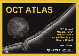
OCT Atlas
Autor SPS Grewal, Manpreet Brar, Mansi Sharma, Mangat Ram Dogra, Dilraj S Grewalen Limba Engleză Hardback – 29 noi 2021
This atlas provides ophthalmologists and trainees with a collection of OCT images to help with the identification, diagnosis and subsequent treatment of common retinal and anterior segment disorders.
The images are compiled from the authors’ own collections using Plex Elite and Cirrus 6000 technology. Fundus angiography images assist with the understanding of related pathologies.
Divided into two sections, the book begins with images illustrating the normal fundus, then numerous different retinal disorders including diabetic retinopathy, macular disorders, retinal detachment, uveitis and toxicities.
Section two covers anterior segment disorders, beginning with images of the normal cornea, then illustrating a range of disorders including corneal dystrophies, ocular surface disorders, keratoconus, glaucoma, and trauma.
Each section features a multitude of images, each with brief descriptive text.
Preț: 1253.20 lei
Preț vechi: 1617.67 lei
-23% Nou
239.83€ • 249.46$ • 197.99£
Carte indisponibilă temporar
Specificații
ISBN-10: 9354650929
Pagini: 220
Dimensiuni: 241 x 330 mm
Greutate: 2.83 kg
Editura: Jp Medical Ltd
Colecția Jaypee Brothers Medical Publishers
Locul publicării:Delhi, India
Cuprins
Section 1: Retina
- Normal OCT
- Vitreous
- Diabetic Retinopathy
- Age-Related Macular Degeneration
- Retinal Vascular Disorder
- Retinal Detachment
- Central Serious Chorioretinopathy
- Macular Holes
- Trauma
- Toxicities
- Macular Dystrophies and Retinal Degenerations
- Pathological Myopia
- Uveitis
- Miscellaneous
- OCT Angiography
Section 2: Anterior Segment
- Normal Cornea
- Corneal Dystrophies and Degenerations
- Ocular Surface Disorders
- Keratitis
- Trauma
- Keratoconus and Corneal Ectasias
- Corneal and Refractive Surgeries
- Optic Nerve Head and RNFL
- OCT Angiography and Glaucoma
- AS-OCT in Glaucoma
Notă biografică
SPS Grewal MBBS MD
CEO
Manpreet Brar MBBS MS
Senior Consultant
Mansi Sharma MBBS DNB FAICO
Consultant
Mangat Ram Dogra MBBS MS
Director
All at Department of Vitreo-retinal Diseases and Surgery, Grewal Eye Institute, Chandigarh, India
Dilraj S Grewal MBBS MD
Associate Professor of Ophthalmology, Vitreoretinal Surgery and Uveitis, Duke Eye Centre, Duke University Medical Centre, Durham, North Carolina, USA
Descriere
Optical coherence tomography (OCT) is a non-invasive imaging test that uses light waves to take cross-sectional pictures of the retina, the light-sensitive tissue lining the back of the eye (eyeSmart). The technique is recognised worldwide as an essential device for diagnosis, assessment and follow up of retinal diseases and glaucoma.
This atlas provides ophthalmologists and trainees with a collection of OCT images to help with the identification, diagnosis and subsequent treatment of common retinal and anterior segment disorders.
The images are compiled from the authors’ own collections using Plex Elite and Cirrus 6000 technology. Fundus angiography images assist with the understanding of related pathologies.
Divided into two sections, the book begins with images illustrating the normal fundus, then numerous different retinal disorders including diabetic retinopathy, macular disorders, retinal detachment, uveitis and toxicities.
Section two covers anterior segment disorders, beginning with images of the normal cornea, then illustrating a range of disorders including corneal dystrophies, ocular surface disorders, keratoconus, glaucoma, and trauma.
Each section features a multitude of images, each with brief descriptive text.
