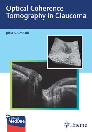
Optical Coherence Tomography in Glaucoma
Autor J Rosdahlen Limba Engleză Hardback – 3 mai 2022
Optical coherence tomography (OCT) is a noninvasive diagnostic imaging modality that enables ophthalmologists to visualize different layers of the optic nerve and retinal nerve fiber layer (RNFL) with astounding detail. Today, OCT is an instrumental tool for screening, diagnosing, and tracking the progression of glaucoma in patients. Optical Coherence Tomography in Glaucoma by renowned glaucoma specialist Jullia A. Rosdahl and esteemed contributors is a one-stop, unique resource that summarizes the clinical utility of this imaging technology, from basics to advanced analyses.
The book features 14 chapters, starting with introductory chapters that discuss development of OCT and its applications for visualizing the optic nerve and macula. In chapter 5, case studies illustrate OCT imaging of the optic nerve, RNFL, and macula in all stages of glaucoma, from patients at risk to those with mild, moderate, and severe diseases. The next three chapters cover the intrinsic relationship between optic nerve structure and function, the use of structure-function maps with examples of their relationship, a comparison of commonly used devices, and artifacts. Subsequent chapters discuss anterior segment OCT and special considerations in pediatric glaucomas and in patients with high refractive errors. The final chapters feature OCT innovations on the horizon, including angiography, swept-source, and artificial intelligence.
Key Highlights
Illustrative case examples provide firsthand clinical insights on how OCT can be leveraged to inform glaucoma treatment
In-depth guidance on recognizing and managing artifacts including case examples and key technical steps to help prevent their occurrence
Pearls on the use of OCT for less common patient scenarios such as pediatric glaucomas and high refractive errors
Future OCT directions including angiography, swept-source, and the use of artificial intelligence
This practical resource is essential reading for ophthalmology trainees and ophthalmologists new to using OCT for glaucoma. The pearls, examples, and novel topics in this book will also help experienced clinicians deepen their knowledge and increase confidence using OCT in daily practice.
This book includes complimentary access to a digital copy on https://medone.thieme.com
Preț: 772.73 lei
Preț vechi: 813.39 lei
-5% Nou
147.88€ • 160.58$ • 124.22£
Carte indisponibilă temporar
Specificații
ISBN-10: 1684202477
Pagini: 212
Ilustrații: 225 Abbildungen
Dimensiuni: 186 x 258 x 17 mm
Greutate: 0.71 kg
Editura: MM – Thieme
Descriere
A comprehensive and user-friendly guide on leveraging OCT for the management of glaucoma
Optical coherence tomography (OCT) is a noninvasive diagnostic imaging modality that enables ophthalmologists to visualize different layers of the optic nerve and retinal nerve fiber layer (RNFL) with astounding detail. Today, OCT is an instrumental tool for screening, diagnosing, and tracking the progression of glaucoma in patients. Optical Coherence Tomography in Glaucoma by renowned glaucoma specialist Jullia A. Rosdahl and esteemed contributors is a one-stop, unique resource that summarizes the clinical utility of this imaging technology, from basics to advanced analyses.
The book features 14 chapters, starting with introductory chapters that discuss development of OCT and its applications for visualizing the optic nerve and macula. In chapter 5, case studies illustrate OCT imaging of the optic nerve, RNFL, and macula in all stages of glaucoma, from patients at risk to those with mild, moderate, and severe diseases. The next three chapters cover the intrinsic relationship between optic nerve structure and function, the use of structure-function maps with examples of their relationship, a comparison of commonly used devices, and artifacts. Subsequent chapters discuss anterior segment OCT and special considerations in pediatric glaucomas and in patients with high refractive errors. The final chapters feature OCT innovations on the horizon, including angiography, swept-source, and artificial intelligence.
Key Highlights
Illustrative case examples provide firsthand clinical insights on how OCT can be leveraged to inform glaucoma treatment
In-depth guidance on recognizing and managing artifacts including case examples and key technical steps to help prevent their occurrence
Pearls on the use of OCT for less common patient scenarios such as pediatric glaucomas and high refractive errors
Future OCT directions including angiography, swept-source, and the use of artificial intelligence
This practical resource is essential reading for ophthalmology trainees and ophthalmologists new to using OCT for glaucoma. The pearls, examples, and novel topics in this book will also help experienced clinicians deepen their knowledge and increase confidence using OCT in daily practice.
This book includes complimentary access to a digital copy on https://medone.thieme.com
Cuprins
2 Development of Optical Coherence Tomography
3 Optical Coherence Tomography of the Optic Nerve
4 Optical Coherence Tomography of the Macula
5 Illustrative Case Examples
6 Structure-Function Relationship
7 Comparison of Common Devices
8 Artifacts and Masqueraders
9 Anterior Segment Optical Coherence Tomography in Glaucoma
10 Special Considerations: OCT in Childhood Glaucomas
11 Special Considerations: High Refractive Errors
12 Future Directions: Optical Coherence Tomography Angiography for Glaucoma
13 Future Directions: Swept-Source OCT for Glaucoma
14 Future Directions: Artificial Intelligence Applications
