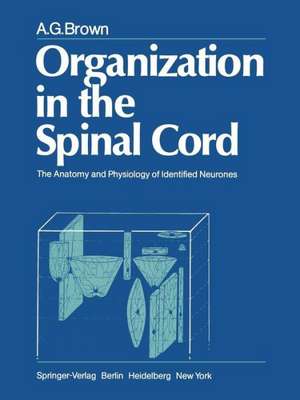
Organization in the Spinal Cord: The Anatomy and Physiology of Identified Neurones
Autor A. G. Brownen Limba Engleză Paperback – 17 noi 2011
Preț: 646.43 lei
Preț vechi: 760.50 lei
-15% Nou
Puncte Express: 970
Preț estimativ în valută:
123.71€ • 134.33$ • 103.92£
123.71€ • 134.33$ • 103.92£
Carte tipărită la comandă
Livrare economică 22 aprilie-06 mai
Preluare comenzi: 021 569.72.76
Specificații
ISBN-13: 9781447113072
ISBN-10: 1447113071
Pagini: 256
Ilustrații: XII, 238 p.
Dimensiuni: 210 x 280 x 13 mm
Greutate: 0.59 kg
Ediția:Softcover reprint of the original 1st ed. 1981
Editura: SPRINGER LONDON
Colecția Springer
Locul publicării:London, United Kingdom
ISBN-10: 1447113071
Pagini: 256
Ilustrații: XII, 238 p.
Dimensiuni: 210 x 280 x 13 mm
Greutate: 0.59 kg
Ediția:Softcover reprint of the original 1st ed. 1981
Editura: SPRINGER LONDON
Colecția Springer
Locul publicării:London, United Kingdom
Public țintă
ResearchCuprins
1 Spinal cord organization: an introduction.- A. Cytoarchitectonic organization of the spinal cord: Rexed’s scheme.- B. An assessment of spinal cord cytoarchitectonics.- C. Physiological classifications of dorsal horn neurones.- D. Resume of spinal cord anatomy.- I. Primary afferent fibres.- 1. Entrance into the spinal cord.- 2. Collaterals.- II. Identified neurones in the spinal cord.- 1. Cells of origin of ascending pathways.- a) Spinocervical tract.- b) Post-synaptic dorsal column pathway.- c) Spinothalamic tract.- d) Dorsal spinocerebellar tract.- e) Ventral spinocerebellar tract.- f) Other ascending tracts.- 2. Identified interneurones with short axons.- a) Renshaw cells.- b) la inhibitory interneurones.- 3. Motoneurones.- 2 Axons innervating hair follicle receptors.- A. Physiology of hair follicle afferent units.- I. Classification.- II. Responses to hair movement.- III. Receptive fields.- B. Central projections of hair follicle afferent fibres.- C. Morphology of axons innervating hair follicle receptors.- I. Entry of axons into the spinal cord, branching and collateral distribution.- II. Morphology of collaterals and their arborizations.- III. Synaptic boutons.- 1. Morphology.- 2. Distribution.- 3. Density.- IV. Organization of collaterals in the dorsal horn.- 1. Organization of collaterals from a single axon.- 2. Somatotopic organization.- 3 Axons innervating rapidly adapting mechanoreceptors in glabrous skin.- A. Axons innervating Pacinian corpuscles.- I. Pacinian corpuscles and their response properties.- II. Central projections of afferent fibres innervating Pacinian corpuscles.- III. Morphology of axons innervating Pacinian corpuscles in the foot and toe pads.- 1. Entry of axons into the spinal cord, branching and collateral distribution.- 2. Morphology ofcollaterals and their arborizations.- 3. Synaptic boutons.- a) Morphology and distribution.- b) Density.- 4. Organization of collaterals in the dorsal horn.- B. Axons innervating rapidly adapting mechanoreceptors in glabrous skin.- I. The receptor and its response properties.- II. Central projections of afferent fibres innervating rapidly adapting mechanoreceptors in the foot and toe pads.- III. Morphology of axons innervating rapidly adapting mechanoreceptors.- 1. Entry of axons into the spinal cord, branching and collateral distribution.- 2. Morphology of collaterals and their arborizations.- 3. Synaptic boutons.- 4. Organization of collaterals in the spinal cord.- 4 Axons innervating slowly adapting Type I mechanoreceptors.- A. Morphology of the Type I receptor.- I. In hairy skin.- II. In glabrous skin.- B. Physiology of Type I units.- I. From hairy skin.- II. From glabrous skin.- C. Central projections of Type I afferent fibres.- D. Morphology of axons innervating Type I receptors.- I. Entry of axons into the spinal cord, branching and collateral distribution.- II. Morphology of collaterals and their arborizations.- III. Synaptic boutons.- 1. Morphology.- 2. Distribution.- 3. Density.- IV. Organization of collaterals in the dorsal horn.- 5 Axons innervating slowly adapting Type II mechanoreceptors.- A. Morphology of the Type II receptor.- B. Physiology of Type II units.- C. Central projections of Type II afferent fibres.- D. Morphology of axons innervating Type II receptors.- I. Entry of axons into the spinal cord, branching and collateral distribution.- II. Morphology of collaterals and their arborizations.- III. Synaptic boutons.- 1. Lamina III and dorsal lamina IV.- 2. Ventral lamina IV.- 3. Lamina V and dorsal lamina VI.- IV. Organization of collaterals in thedorsal horn.- 6 Spinocervical tract neurones.- A. Physiology of the spinocervical tract.- I. Types of unit in the tract.- II. Actions of descending control systems.- III. Transmission of information through the tract.- B. Anatomy of the spinocervical tract.- I. Location of neurones.- II. Density and distribution of neurones.- III. Somatotopic organization.- IV. Morphology of neurones.- 1. Somata and dendritic trees.- 2. Dendritic spines.- 3. Axons and axon collaterals.- 4. Synaptic boutons.- C. Relationships between the anatomy and the physiology of spinocervical tract neurons.- I. Receptive field position and dendritic trees.- II. Receptor input and morphology.- 1. Dendritic trees.- 2. Axon collaterals.- 3. Relationships between dendritic trees and receptive fields of adjacent neurons.- 4. Relationships between dendritic trees and hair follicle afferent fibre collaterals.- 7 Relationships between hair follicle afferent fibres and spinocervical tract neurones.- A. Anatomy of the relationships between hair follicle afferent arborizations and spinocervical tract neurones.- I. Relationships where the afferent fibre and the neurone had receptive fields on different areas of skin: the negative results.- II. Relationships where the receptive field of the neurone contained the field of the afferent fibre: the positive results.- Positions of terminal arborizations of afferent fibres and dendritic trees of neurones.- Number of afferent collaterals distributed to each cell…..- 3. Contacts between afferent fibres and neurones.- a.) Numbers and positions of synapses and their relationships to receptive field positions.- b.) Types and arrangements of synaptic contacts.- 4. Conclusions.- 8 Neurones with axons ascending the dorsal columns.- A. Physiology of neurones with axonsascending the dorsal columns.- I. Response properties.- II. Input from non-primary sources.- III. Axonal conduction velocities.- B. Anatomy of neurones with axons ascending the dorsal columns...- I. Location.- II. Density.- III. Cellular anatomy.- 1. Size of cell bodies.- 2. Dendritic trees.- 3. Axonal projections.- 4. Axon collaterals.- C. Organization of the post-synaptic dorsal column pathway.- I. Correlations between form and function of the neurones…..- II. Comparison with the spinocervical tract.- 9 Other dorsal horn neurones.- A. Dorsal horn neurones with unidentified axonal projections.- I. Neurones with somata in lamina III.- II. Neurones with somata in lamina IV.- III. Neurones with somata ventral to lamina IV.- IV. Conclusions.- 10 The organization of the dorsal horn.- A. Organization of input from the skin.- I. Segregation of input according to axonal diameter and afferent unit type.- II. Specificity of the morphology of axon collateral arborizations according to afferent unit type.- III. Receptive field transformation.- IV. Somatotopic organization.- V. Collateral spacing.- B. The neurones of the dorsal horn.- I. Lamina I (the postero-marginal cell layer).- 1. Anatomy.- a) Dendritic organization.- b) Axonal projections.- 2. Physiological properties.- II. Lamina II (the substantia gelatinosa).- 1. Anatomy.- 2. Physiology.- III. Laminae III-VI.- 1. Anatomy.- a) Dendritic trees..- b) Axonal projections.- c) Somatotopic organization.- C. Descending input to the dorsal horn.- I. Descending pathways: actions and terminations.- 1. Corticospinal tract.- 2. Raphe spinal system.- 3. Reticulospinal pathways.- 4. Other descending systems.- II. Summary.- 11 Afferent fibres from primary endings in muscle spindles.- A. Central projections of muscle spindle primaryendings.- B. Morphology of Group la afferent fibres.- I. Entry of axons into the spinal cord, branching and collateral distribution.- II. Morphology of collaterals.- III. Terminal arborizations.- 1. In the intermediate region (lamina VI).- 2. In the la inhibitory interneurone region (lamina VII).- 3. In the motor nuclei (lamina IX).- IV. Synaptic boutons.- 1. In the intermediate region (lamina VI).- 2. In the la inhibitory interneurone region (lamina VII).- 3. In the motor nuclei (lamina IX).- V. Organization of collaterals.- 12 Afferent fibres from Golgi tendon organs.- A. Central projections of afferent fibres from tendon organs.- I. Afferent fibres from tendon organs.- II. Central effects of lb input: autogenetic inhibition.- III. Location of interneurones excited by lb afferent fibres.- B. Morphology of axons innervating Golgi tendon organs.- I. Entry of axons into the spinal cord, branching and collateral distribution.- II. Morphology of lb collaterals.- III. Terminal arborizations and synaptic boutons of collaterals...- IV. Organization of collaterals.- 13 Afferent fibres from secondary endings in muscle spindles.- A. Central projections of axons from muscle spindle secondary endings.- I. Terminations of Group II fibres.- II. Location of neurones activated by Group II axons.- III. Central effects of impulses in Group II fibres.- B. Morphology of axons innervating muscle spindle secondary endings.- I. Entry of axons into the spinal cord, branching and collateral distribution.- II. Morphology of collaterals.- III. Terminal arborizations and synaptic boutons.- IV. Organization of collaterals.- 14 Relationships between Group la afferent fibres and motoneurones.- A. ?-Motoneurones.- I. Anatomy.- II. Electrophysiology.- B. Actions of la afferent fibres uponmotoneurones.- C. Ia afferent fibre terminations upon motoneurones.- I. Terminations upon motoneuronal somata and proximal dendrites.- II. Terminations upon dendritic trees.- III. Conclusions.- Appendix 1 Methods.- A. Electrophysiological methods.- I. Solutions for intracellular ionophoresis of horseradish peroxidase.- II. Microelectrodes.- III. Intracellular injection of horseradish peroxidase.- IV. Histological methods.- Appendix 2 Nomenclature.- A. Anatomy of the spinal grey matter.- B. Nomenclature for somatosensory neurons.- I. Afferent input.- II. Location of the neuronal soma.- References.


