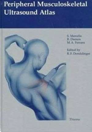
Peripheral Musculoskeletal Ultrasound Atlas
Autor Robert F. Dondelinger, S. Marcelis, B. Daenen, M. A. Ferraraen Limba Engleză Hardback – 23 apr 1996
Straightforward commentary and 750 illustrations - including sonograms and line drawings - combine to make this book an authoritative review of high-definition ultrasonography in diagnosing musculoskeletal pathology of the extremities.
This innovative, applications-oriented guide systematically covers:
This innovative, applications-oriented guide systematically covers:
- State-of-the-art instrumentation and examination techniques, with expert advice on probe positioning
- Common technical problems, diagnostic pitfalls, and useful preventive and corrective actions
- Normal and pathologic ultrasound findings for muscle, tendon, ligament, periosteum and bone, joint capsule, bursa and synovium, cartilage, vessel, nerves, fat, and skin
- Pathologic regional ultrasound findings for the shoulder, arm, elbow, forearm, wrist, hand, hip, thigh, knee, leg, ankle, and foot
- A wide range of specific diagnostic applications, including diagnosis of tendon tears, hematomas, fractures, joint effusions, foreign bodies, and more
Preț: 1038.80 lei
Preț vechi: 1115.81 lei
-7% Nou
Puncte Express: 1558
Preț estimativ în valută:
198.80€ • 207.29$ • 165.22£
198.80€ • 207.29$ • 165.22£
Carte tipărită la comandă
Livrare economică 20-26 martie
Preluare comenzi: 021 569.72.76
Specificații
ISBN-13: 9783131027719
ISBN-10: 3131027711
Pagini: 213
Ilustrații: 750
Dimensiuni: 210 x 297 mm
Greutate: 1.04 kg
Ediția:1st Edition
Editura: Thieme
Colecția Thieme
ISBN-10: 3131027711
Pagini: 213
Ilustrații: 750
Dimensiuni: 210 x 297 mm
Greutate: 1.04 kg
Ediția:1st Edition
Editura: Thieme
Colecția Thieme
Textul de pe ultima copertă
Straightforward commentary and 750 illustrations - including sonograms and line drawings - combine to make this book an authoritative review of high-definition ultrasonography in diagnosing musculoskeletal pathology of the extremities.
This innovative, applications-oriented guide systematically covers:
This innovative, applications-oriented guide systematically covers:
- State-of-the-art instrumentation and examination techniques, with expert advice on probe positioning
- Common technical problems, diagnostic pitfalls, and useful preventive and corrective actions
- Normal and pathologic ultrasound findings for muscle, tendon, ligament, periosteum and bone, joint capsule, bursa and synovium, cartilage, vessel, nerves, fat, and skin
- Pathologic regional ultrasound findings for the shoulder, arm, elbow, forearm, wrist, hand, hip, thigh, knee, leg, ankle, and foot
- A wide range of specific diagnostic applications, including diagnosis of tendon tears, hematomas, fractures, joint effusions, foreign bodies, and more
Descriere
The authoritative review of the use of high-definition ultrasonography in diagnosing musculoskeletal pathology of the extremities. The first part, organized according to tissue type, describes their typical appearances of normal tissues and the general appearances of pathologic changes. The second part, organized according to anatomic region, describes the ultrasound findings, normal structures, as well as the classic and rare diseases that may affect these regions. Clear guidelines on the correct placement of the scanner, pitfalls, and artifacts are included.
