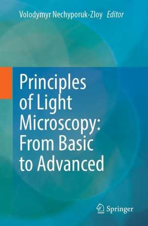
Principles of Light Microscopy: From Basic to Advanced
Editat de Volodymyr Nechyporuk-Zloyen Limba Engleză Paperback – 30 noi 2022
The book covers key microscopy principles and explains the various techniques such as epifluorescence microscopy, confocal/live cell imaging, SIM/light sheet microscopy, and many more. Easy-to-understand protocols provide helpful guidance for practical implementation in various commercially available imaging systems. The reader is introduced to histology and further be guided through advanced image acquisition, classification and analysis.
The book is written by experienced imaging specialists from the UK, other EU countries, the US and Asia, and is based on advanced training courses for master students and PhD students. Readers are not expected to be familiar with imaging and microscopy technologies, but are introduced to the subject step by step. This textbook is indented for biomedical and medical students, as well as scientists and postdocs who want to acquire a thorough knowledge of microscopy, or gain a comprehensive overview of modern microscopy techniques used in various research laboratories and imaging facilities.
Chapter 4 is available open access under a Creative Commons Attribution 4.0 International License via link.springer.com.
Preț: 434.02 lei
Preț vechi: 456.87 lei
-5% Nou
Puncte Express: 651
Preț estimativ în valută:
83.06€ • 90.19$ • 69.77£
83.06€ • 90.19$ • 69.77£
Carte tipărită la comandă
Livrare economică 22 aprilie-06 mai
Preluare comenzi: 021 569.72.76
Specificații
ISBN-13: 9783031044762
ISBN-10: 3031044762
Pagini: 324
Ilustrații: VIII, 324 p. 1 illus.
Dimensiuni: 155 x 235 mm
Greutate: 0.47 kg
Ediția:1st ed. 2022
Editura: Springer International Publishing
Colecția Springer
Locul publicării:Cham, Switzerland
ISBN-10: 3031044762
Pagini: 324
Ilustrații: VIII, 324 p. 1 illus.
Dimensiuni: 155 x 235 mm
Greutate: 0.47 kg
Ediția:1st ed. 2022
Editura: Springer International Publishing
Colecția Springer
Locul publicării:Cham, Switzerland
Cuprins
Chapter 1. The physical principles of light propagation and light-matter interactions.- Chapter 2. Design, alignment and usage of infinity-corrected microscope.- Chapter 3. Epifluorescence Microscopy.- Chapter 4. Basic digital image design, processing, analysis, management, and presentation.- Chapter 5. Confocal Microscopy.- Chapter 6. Live-cell Imaging: A Balancing Act Between Speed, Sensitivity and Resolution.- Chapter 7. Structured Illumination Microscopy.- Chapter 8. Stimulated Emission Depletion Microscopy.- Chapter 9. Two-Photon Imaging.- Chapter 10. Modern microscopy image analysis: quantifying colocalization on a mobile device.- Chapter 11. Spinning Disk Microscopy.- Chapter 12. Light Sheet Fluorescence Microscopy.- Chapter 13. Localization microscopy: a review of the progress in methods and applications.
Notă biografică
Volodymyr Nechyporuk Zloy, PhD, is Bio Imaging Lab Manager at Nikon Europe, where his activities encompass the management of the equipment, the development of the new imaging services and education. Volodymyr has developed several microscopy courses for master and PhD students.
Textul de pe ultima copertă
This textbook is an excellent guide to microscopy for students and scientists, who use microscopy as one of their primary research and analysis tool in the laboratory.
The book covers key microscopy principles and explains the various techniques such as epifluorescence microscopy, confocal/live cell imaging, SIM/light sheet microscopy, and many more. Easy-to-understand protocols provide helpful guidance for practical implementation in various commercially available imaging systems. The reader is introduced to histology and further be guided through advanced image acquisition, classification and analysis.
The book is written by experienced imaging specialists from the UK, other EU countries, the US and Asia, and is based on advanced training courses for master students and PhD students. Readers are not expected to be familiar with imaging and microscopy technologies, but are introduced to the subject step by step. This textbook is indented for biomedical and medical students, as well as scientists and postdocs who want to acquire a thorough knowledge of microscopy, or gain a comprehensive overview of modern microscopy techniques used in various research laboratories and imaging facilities.
Chapter 4 is available open access under a Creative Commons Attribution 4.0 International License via link.springer.com.
Caracteristici
Well-structured textbook on imaging techniques Covers basic microscopic principles and explains the various techniques Supplemented with instructions and protocols for practical application
