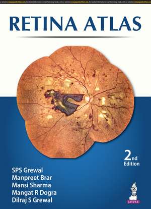
Retina Atlas
Autor SPS Grewal, Manpreet Brar, Mansi Sharma, R Mangat Dogra, S Dilraj Grewalen Limba Engleză Hardback – 11 mar 2024
The images are procured from Eidon scanner technology and also include optical coherence tomography (OCT) pictures to assist with understanding of related pathologies.
Divided into nine sections, the book begins with images illustrating the normal fundus. Each of the following sections covers a different retinal disorder including diabetic retinopathy, macula disorders, retinal detachment, ocular tumours and hereditary diseases.
Each section features a multitude of images, each with brief descriptive text to assist understanding.
The second edition of this illustrative atlas has been fully revised and updated to reflect the latest developments and knowledge in the field.
Previous edition (97893895874432) published in 2020.
Preț: 529.20 lei
Preț vechi: 667.97 lei
-21% Nou
Puncte Express: 794
Preț estimativ în valută:
101.27€ • 105.60$ • 84.17£
101.27€ • 105.60$ • 84.17£
Carte indisponibilă temporar
Doresc să fiu notificat când acest titlu va fi disponibil:
Se trimite...
Preluare comenzi: 021 569.72.76
Specificații
ISBN-13: 9789356968806
ISBN-10: 9356968802
Pagini: 318
Dimensiuni: 241 x 330 x 22 mm
Greutate: 2.1 kg
Ediția:2
Editura: JAYPEE BROTHERS MEDICAL PUBLISHERS PVT LTD
Colecția Jaypee Brothers,Medical Publishers Pvt. Ltd.
Locul publicării:Delhi, India
ISBN-10: 9356968802
Pagini: 318
Dimensiuni: 241 x 330 x 22 mm
Greutate: 2.1 kg
Ediția:2
Editura: JAYPEE BROTHERS MEDICAL PUBLISHERS PVT LTD
Colecția Jaypee Brothers,Medical Publishers Pvt. Ltd.
Locul publicării:Delhi, India
Cuprins
CHAPTER 1: Normal Fundus
CHAPTER 2: Diabetic Retinopathy
CHAPTER 3: Retinal Vascular Disorders
CHAPTER 4: Macula
CHAPTER 5: Retinal Detachment
CHAPTER 6: Inflammatory
CHAPTER 7: Hereditary
CHAPTER 8: Ocular Tumors and Optic Nerve Disorders
CHAPTER 9: Miscellaneous
Glossary
CHAPTER 2: Diabetic Retinopathy
CHAPTER 3: Retinal Vascular Disorders
CHAPTER 4: Macula
CHAPTER 5: Retinal Detachment
CHAPTER 6: Inflammatory
CHAPTER 7: Hereditary
CHAPTER 8: Ocular Tumors and Optic Nerve Disorders
CHAPTER 9: Miscellaneous
Glossary
Notă biografică
SPS Grewal MBBS MD
Manpreet Brar MBBS MS
Mansi Sharma MBBS DNB FAICO
Mangat R Dogra MBBS MS
Dilraj S Grewal MBBS MD
Manpreet Brar MBBS MS
Mansi Sharma MBBS DNB FAICO
Mangat R Dogra MBBS MS
Dilraj S Grewal MBBS MD
Descriere
This atlas provides ophthalmologists with a collection of images to help with the identification, diagnosis and subsequent treatment of retinal disorders. The images are procured from Eidon scanner technology and also include optical coherence tomography (OCT) pictures to assist with understanding of related pathologies. Each section features a multitude of images, each with brief descriptive text to assist understanding.
