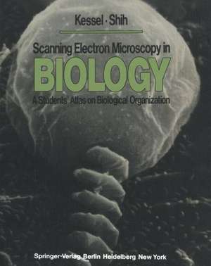
Scanning Electron Microscopy in BIOLOGY: A Students’ Atlas on Biological Organization
Autor R. G. Kessel, C. Y. Shihen Limba Engleză Paperback – 10 ian 2012
Preț: 651.67 lei
Preț vechi: 766.67 lei
-15% Nou
Puncte Express: 978
Preț estimativ în valută:
124.70€ • 135.100$ • 105.15£
124.70€ • 135.100$ • 105.15£
Carte tipărită la comandă
Livrare economică 24 aprilie-08 mai
Preluare comenzi: 021 569.72.76
Specificații
ISBN-13: 9783642808364
ISBN-10: 3642808360
Pagini: 368
Ilustrații: X, 348 p. 160 illus.
Dimensiuni: 203 x 254 x 19 mm
Greutate: 0.73 kg
Ediția:Softcover reprint of the original 1st ed. 1976
Editura: Springer Berlin, Heidelberg
Colecția Springer
Locul publicării:Berlin, Heidelberg, Germany
ISBN-10: 3642808360
Pagini: 368
Ilustrații: X, 348 p. 160 illus.
Dimensiuni: 203 x 254 x 19 mm
Greutate: 0.73 kg
Ediția:Softcover reprint of the original 1st ed. 1976
Editura: Springer Berlin, Heidelberg
Colecția Springer
Locul publicării:Berlin, Heidelberg, Germany
Public țintă
ResearchCuprins
1 Introduction.- Comparison of the Scanning Electron Microscope with Other Microscopes.- Basic Theory and Operation of the Scanning Electron Microscope.- Modes of Operation of the Scanning Electron Microscope.- Methods of Specimen Preparation in Scanning Electron Microscopy.- 2 One-Celled Organisms.- Ciliate Protozoa.- Flagellate Protozoa.- Amoeboid Protozoa.- 3 Cells in Culture.- Surface Specializations.- Variations in Cell Surface Specializations.- Cells in Mitosis.- Chick Embryo Chondroblasts.- Phagocytosis by Normal and Abnormal Tissue Culture Cells.- Induced Morphogenesis of Glial Cells.- 4 Prokaryotes.- Bacteria.- Blue-green Algae.- 5 Fungi and Algae.- Slime Molds.- Fungi.- Algae.- 6 Multicellular Plants.- Liverworts and Mosses.- Lower Vascular Plants.- Gymnosperms.- 7 Organ Systems of Angiosperms.- Roots.- Shoot Apex.- Stem.- Leaf.- Flower.- Seed.- 8 Multicellular Animals.- Sponges.- Hydra and Other Coelenterates.- Flatworms.- Spiny-headed Worms.- Nematodes.- Ectoprocts or Lophophorate Coelomates.- Annelid Worms.- Molluscs.- Arthropods.- 9 Tissue and Organ Systems of Animals.- Blood.- Muscle Tissue.- Digestive System.- Respiratory System.- Excretory System.- Male Reproductive System.- Female Reproductive System.- Sense Organs.- 10 Development.- Fertilization and the Cortical Reaction in Sea Urchins.- Embryology of the Frog.
