Cantitate/Preț
Produs

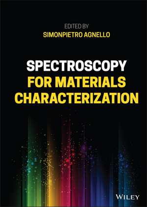
Spectroscopy for Materials Characterization

Autor S Agnello
en Limba Engleză Hardback – 4 noi 2021

SPECTROSCOPY FOR MATERIALS CHARACTERIZATION

Learn foundational and advanced spectroscopy techniques from leading researchers in physics, chemistry, surface science, and nanoscience

In Spectroscopy for Materials Characterization, accomplished researcher Simonpietro Agnello delivers a practical and accessible compilation of various spectroscopy techniques taught and used to today. The book offers a wide-ranging approach taught by leading researchers working in physics, chemistry, surface science, and nanoscience. It is ideal for both new students and advanced researchers studying and working with spectroscopy.

Topics such as confocal and two photon spectroscopy, as well as infrared absorption and Raman and micro-Raman spectroscopy, are discussed, as are thermally stimulated luminescence and spectroscopic studies of radiation effects on optical materials.

Each chapter includes a basic introduction to the theory necessary to understand a specific technique, details about the characteristic instrumental features and apparatuses used, including tips for the appropriate arrangement of a typical experiment, and a reproducible case study that shows the discussed techniques used in a real laboratory.

Readers will benefit from the inclusion of:

  • Complete and practical case studies at the conclusion of each chapter to highlight the concepts and techniques discussed in the material
  • Citations of additional resources ideal for further study
  • A thorough introduction to the basic aspects of radiation matter interaction in the visible-ultraviolet range and the fundamentals of absorption and emission
  • A rigorous exploration of time resolved spectroscopy at the nanosecond and femtosecond intervals

Perfect for Master and Ph.D. students and researchers in physics, chemistry, engineering, and biology, Spectroscopy for Materials Characterization will also earn a place in the libraries of materials science researchers and students seeking a one-stop reference to basic and advanced spectroscopy techniques.

Citește tot Restrânge

Preț: 101628 lei

Preț vechi: 111680 lei
-9% Nou

Puncte Express: 1524

Preț estimativ în valută:
19455 20009$ 16140£

Carte tipărită la comandă

Livrare economică 20 februarie-06 martie

Preluare comenzi: 021 569.72.76

Specificații

ISBN-13: 9781119697329
ISBN-10: 1119697328
Pagini: 496
Dimensiuni: 186 x 260 x 32 mm
Greutate: 1.06 kg
Editura: Wiley
Locul publicării:Hoboken, United States

Cuprins

Preface xv List of Contributors xvii 1 Radiation-Matter Interaction Principles: Optical Absorption and Emission in the Visible-Ultraviolet Region 1 Simonpietro Agnello 1.1 Empirical Aspects of Radiation-Matter Interaction 1 1.1.1 Optical Absorption: The Lambert-Beer Law 1 1.1.2 Emission: Fluorescence and Phosphorescence 5 1.2 Microscopic Point of View 7 1.2.1 Einstein Coefficients 7 1.2.2 Oscillator Strength, Lifetime, Quantum Yield 11 1.2.3 Vibronic States: Homogeneous and Inhomogeneous Lineshape 14 1.2.4 Jablonski Energy Level Diagram: Permitted and Forbidden Transitions 20 1.2.5 Excited States Rate Equations 22 1.3 Instrumental Setups 23 1.3.1 Typical Block Diagram of Spectrometers 23 1.3.2 Light Sources 24 1.3.3 Dispersion Elements: Gratings and Resolution Power 25 1.3.4 Detectors: Photodiode, Photomultiplier, Charge Coupled Device 27 1.4 Case Studies 29 1.4.1 Optical Absorption in Visible-Ultraviolet Range 29 1.4.1.1 Scanning Device (Bandwidth and Scanning Speed Effects) 29 1.4.1.2 CCD Fiber Optic Device 31 1.4.2 Photoluminescence 31 1.4.2.1 Emission and Excitation Spectra: Energy Levels Reconstruction 32 References 33 2 Time-Resolved Photoluminescence 35 Marco Cannas and Lavinia Vaccaro 2.1 Introduction to Photoluminescence Spectroscopy 35 2.1.1 Photoluminescence Properties Related to Points Defects: Electron-Phonon Coupling 35 2.1.2 Optical Transitions: The Franck-Condon Principle 38 2.1.3 Zero-Phonon Line 40 2.1.4 Phonon Line Structure 43 2.1.5 Vibrational Structure 45 2.1.6 Inhomogeneous Effects 48 2.2 Experimental Methods and Analysis 48 2.2.1 Time-Resolved Luminescence 48 2.2.2 Site-Selective Luminescence 50 2.2.3 Basic Design of Experimental Setup: Pulsed Laser Sources; Monochromators; Detectors 51 2.2.3.1 Tunable Laser 52 2.2.3.2 Time-Resolved Detection System: Spectrograph and Intensified CCD Camera 52 2.3 Case Studies: Luminescent Point Defects in Amorphous SiO2 54 2.3.1 Emission Spectra and Lifetime Measurements 55 2.3.2 Zero-Phonon Line Probed by Site-Selective Luminescence 58 References 63 3 Ultrafast Optical Spectroscopies 65 Alice Sciortino and Fabrizio Messina 3.1 Femtosecond Spectroscopy: An Overview 65 3.2 Ultrafast Optical Pulses 67 3.2.1 General Properties 67 3.2.1.1 Dispersion Effect: Group Velocity Dispersion 67 3.2.2 Nonlinear Optics: Basis and Applications 69 3.2.2.1 Second Harmonic Generation and Sum Frequency Generation 69 3.2.2.2 Noncollinear Optical Parametric Amplifier 70 3.2.2.3 Supercontinuum Generation 72 3.3 Transient Absorption Spectroscopy 73 3.3.1 The Experimental Method 74 3.3.2 Typical Experimental Setups 76 3.3.3 Data Analysis and Interpretation 78 3.4 Ultrafast Fluorescence Spectroscopies 79 3.4.1 FLUC: The Experimental Method 80 3.4.2 FLUC: Typical Experimental Setups 80 3.4.3 FLUC: Data Analysis and Interpretation 82 3.4.4 Kerr-Based Femtosecond Fluorescence Spectroscopy 82 3.5 Femtosecond Stimulated Raman Spectroscopy 83 3.5.1 The Experimental Method 83 3.5.2 Typical Experimental Setups 84 3.5.3 Data Analysis and Interpretation 87 3.6 Case Studies 88 3.6.1 Ultrafast Relaxation Dynamics of Molecules in Solution Phase 88 3.6.2 Relaxation of Excited Charge Carriers and Excitons in Semiconductor Nanoparticles 89 3.6.3 Ultrafast Relaxation Dynamics of Carbon-based Nanomaterials 91 References 92 4 Confocal and Two-Photon Spectroscopy 97 Giuseppe Sancataldo and Valeria Vetri 4.1 Introduction and Historical Perspectives 97 4.1.1 Point Spread Function and Optical Resolution 98 4.1.2 Optical Sectioning and Imaging of 3D Samples 101 4.2 Fluorescence Imaging 102 4.2.1 Laser Scanning Confocal Fluorescence Microscope 103 4.2.2 Two-Photon Microscope 105 4.2.3 The Importance of Sample Preparation from Solid State to Dynamic Specimens 108 4.2.4 Setting Up a Measurement 109 4.3 Spectroscopy Using a Microscope 110 4.3.1 Observables in Fluorescence Microscopy 111 4.3.2 Measuring Dynamics: Gaining Information Below Resolution 113 4.4 Case Studies 117 4.4.1 Understanding Microstructures and Mechanistic Aspects in Materials 117 4.4.2 Fluctuation Methods for the Analysis of Nanosystems 121 References 124 5 Infrared Absorption Spectroscopy 129 Tiziana Fiore and Claudia Pellerito 5.1 Fundamentals 129 5.1.1 Introduction 130 5.1.2 Basic Principles 130 5.1.3 Infrared Spectra 135 5.1.4 Fourier Transform Infrared Spectrometers (Interferometers) 137 5.2 Sources and Detectors 140 5.3 Techniques and Sampling Methods 144 5.3.1 Transmission Methods 144 5.3.1.1 Solid Samples 144 5.3.1.2 Liquid and Solution Samples 147 5.3.1.3 Gas Samples 148 5.3.2 Attenuated Total Reflectance (ATR) Method 148 5.3.3 FTIR Microspectroscopy 150 5.3.4 AFM-IR Spectroscopy 150 5.3.5 Hyphenated Techniques 150 5.4 Applications and Case Studies 151 5.4.1 Chemical Characterization and Kinetics 151 5.4.2 Surfaces 152 5.4.3 Medical and Life Science (Pharmaceutical, Medical, Biological, Biotechnological) 153 5.4.4 Cultural Heritage and Forensic 156 5.4.5 Environmental and Geological 157 5.4.6 Food Industry 158 References 158 6 Raman and Micro-Raman Spectroscopy 169 Giuliana Faggio, Rossella Grillo, and Giacomo Messina 6.1 Basic Theory 169 6.1.1 Introduction 169 6.1.2 Spectroscopic Units 169 6.1.3 Molecular Vibrations 170 6.1.4 Classical Theory of the Raman Scattering 171 6.1.5 Simplified Quantum Approach to Raman Scattering 174 6.1.6 Raman and IR Activities 178 6.1.7 Crystal Vibrations 180 6.1.8 Raman Scattering in Crystals 183 6.1.9 Surface-Enhanced Raman Scattering (SERS) 185 6.2 Instrumentation 187 6.2.1 Laser Sources and Optical Filters 187 6.2.2 Monochromators 188 6.2.3 Detectors 189 6.2.4 Raman Microscopy and Raman Mapping 189 6.3 Case Studies 191 6.3.1 Raman Indicators 191 6.3.2 Identification of Materials and Crystalline Quality 191 6.3.3 Graphene and Graphite Raman Spectra 193 6.3.4 Doping Detection 196 6.3.5 Basic Examples of SERS 196 References 198 7 Thermally Stimulated Luminescence 201 Federico Moretti 7.1 Theory of Thermally Stimulated Luminescence 202 7.1.1 Simple Model 205 7.1.1.1 First-Order Kinetics 207 7.1.1.2 Second-Order Kinetics 211 7.1.1.3 General-Order Kinetics 211 7.1.2 Localized Transitions 213 7.1.3 Beyond the Ideal Behavior 214 7.1.3.1 Luminescence Quenching 215 7.1.3.2 Trap Energy Distributions 216 7.2 Data Analysis Methods 216 7.2.1 Initial Rise 217 7.2.2 Peak Shape 218 7.2.3 Heating Rate Method 220 7.2.4 Glow Curve Fit 221 7.3 Instrumentation and Considerations on Samples 221 7.4 Case Studies 222 7.4.1 Lanthanide Energy Level Position in the Bandgap 223 7.4.2 Bandgap Engineering 224 7.4.3 Correlation of TSL Data with EPR Results 225 Note 225 References 226 8 Spectroscopic Studies of Radiation Effects on Optical Materials 229 Sylvain Girard, Vincenzo De Michele, and Adriana Morana 8.1 Introduction 229 8.1.1 Radiation Environments 229 8.1.2 Applications for Optical Materials 230 8.2 Radiation-Induced Effects on Optical Materials and Optical Fibers 231 8.2.1 Radiation-Induced Attenuation - RIA 231 8.2.2 Radiation-Induced Emission - RIE 233 8.2.3 Radiation-Induced Compaction - RIC and Refractive Index Change - RIRIC 234 8.2.4 Origins of Radiation-Induced Optical Changes 234 8.3 Radiation-Induced Attenuation Measurements 235 8.3.1 Postirradiation RIA Measurements 235 8.3.1.1 Bulk Glasses 235 8.3.1.2 Optical Fibers 235 8.3.2 In Situ RIA Measurements 236 8.3.2.1 Bulk Glasses 236 8.3.2.2 Optical Fibers 237 8.3.3 Exploitation of RIA Spectra: Point Defect Identification 241 8.3.3.1 Spectral Decomposition 241 8.3.3.2 Point Defect Kinetics 243 8.4 Radiation-Induced Luminescence (RIL) 243 8.4.1 Architectures of Fiber-Based Sensors: Extrinsic and Intrinsic 243 8.4.2 Calibration of the RIL Versus Proton Flux 245 8.4.3 Bragg Peak Measurements for Proton-Therapy Applications 245 8.5 Case Studies 246 8.5.1 Characterization of Bulk Glasses for Space Optical Systems 246 8.5.2 Fiber-Based Dosimetry with Phosphorus-Doped Optical Fibers 247 8.5.3 Proton Flux Measurements Through the RIL of Optical Fibers 249 References 249 9 Electron Paramagnetic Resonance Spectroscopy (EPR) 253 Antonino Alessi and Franco Gelardi 9.1 Introduction 253 9.2 Basic Principle of EPR 253 9.3 Anisotropy of g and Spectral Lineshape 255 9.4 The EPR Lineshape in Powder or in Amorphous 257 9.5 Hyperfine Interactions 258 9.6 Paramagnetic Center with S = 1 261 9.7 Basics of Continuous Wave EPR Setup 263 9.8 Parameters for EPR Signal Acquisition 266 9.9 Cw EPR Case Studies 268 9.10 Time-Resolved EPR Spectroscopy 270 9.10.1 Saturation Transients 270 9.10.2 Spin Nutations 272 9.10.3 Free Induction Decay 274 9.10.4 Spin Echo 276 References 277 10 Nuclear Magnetic Resonance Spectroscopy 281 Alberto Spinella and Pellegrino Conte 10.1 Introduction 281 10.2 NMR General Concepts 281 10.2.1 Nuclear Spin and Magnetic Moment 281 10.2.2 Spin Precession and Larmor Frequency 283 10.2.3 Longitudinal Magnetization 283 10.2.4 Transverse Magnetization and NMR Signal 284 10.2.5 Spin Interactions 285 10.2.6 Fourier Transform NMR 287 10.3 Liquid-State NMR 288 10.3.1 The NMR Spectrometer 288 10.3.2 Sample Preparation 288 10.3.3 How to Set an Experiment 289 10.3.4 Longitudinal Relaxation Time Measurement 289 10.3.5 Transverse Relaxation Time Measurement 290 10.3.6 2D-Liquid-State NMR Techniques 291 10.3.7 Considerations on the Molecular Dynamics by NMR Spectroscopy 292 10.4 Solid-State NMR 293 10.4.1 Powdered Samples 293 10.4.2 Cross-Polarization and Heteronuclear Decoupling 294 10.4.3 Magic-Angle Spinning 296 10.4.4 Homonuclear Dipolar Decoupling 299 10.4.5 2D-Solid State NMR Techniques 299 10.4.6 Recoupling Techniques 300 10.4.7 Molecular Dynamics by Solid-State NMR Spectroscopy 301 10.5 Nonconventional NMR Techniques 301 10.5.1 Time Domain NMR 302 10.5.2 Fast Field Cycling NMR Relaxometry 302 10.5.3 Earth's Magnetic Field NMR 309 10.6 Case Studies 309 10.6.1 Polymers and Polymer-Based Composites 309 10.6.2 Mesoporous Materials 310 10.6.3 Cultural Heritage 311 10.6.4 Food 313 10.6.5 Environmental NMR: Rocks, Soils, Waters, Air 313 10.6.6 NMR of "Exotic" Nuclei 314 References 315 11 X-Ray Absorption Spectroscopy and X-Ray Raman Scattering Spectroscopy for Energy Applications 319 Alessandro Longo, Francesco Giannici, and Christoph J. Sahle 11.1 Introduction 319 11.2 The X-Ray Absorption Coefficient and the EXAFS Technique 320 11.2.1 The EXAFS Equation and the Key Approximations 322 11.2.1.1 Many-Body Effects 323 11.2.1.2 Inelastic Effects 324 11.2.2 Multiple Scattering Theory: Basic Information 325 11.2.3 XANES or Near-Edge X-Ray Absorption Fine Structure and Pre-Edge Region 328 11.3 EXAFS: Data Analysis Overview 331 11.4 Experimental Setups 333 11.4.1 Transmission Geometry 333 11.4.2 Fluorescence Geometry 334 11.5 X-Ray Raman Scattering Spectroscopy 335 11.5.1 Theoretical Background 335 11.5.2 Experimental Setup 338 11.5.2.1 Instrumentation 338 11.5.2.2 Data Processing 338 11.6 Case Studies: Application of XAFS and XRS for Energy Materials 339 11.6.1 CO Oxidation Reaction: The Au/CeO2 Catalyst 339 11.6.2 Materials for Solid Oxide Fuel Cells 340 11.6.3 Oxide-Ion Conductors: Dopants and Vacancies 342 11.6.4 Proton-Conducting Oxides 343 11.6.5 The Role of Oxygen in Fuel Cell Cathodes 344 References 346 12 X-Ray Photoelectron Spectroscopy 351 Michelangelo Scopelliti 12.1 General Principles 351 12.2 Instrumental Setup 352 12.2.1 Vacuum and Ultrahigh Vacuum, UHV 353 12.2.1.1 Roughing Pumps 354 12.2.1.2 Turbomolecular Pumps 355 12.2.1.3 Ion Pumps 355 12.2.1.4 Titanium Sublimation Pumps 356 12.2.2 Magnetic Shielding 356 12.2.3 Sources 356 12.2.4 Sample Manipulators 358 12.2.5 Charge Neutralization Systems 359 12.2.5.1 Electron Guns 360 12.2.5.2 Ion Guns 360 12.2.6 Analyzers and Detectors 361 12.3 Applications 362 12.3.1 Quantitative Analysis 364 12.3.2 Qualitative Analysis 365 12.3.3 Surface Maps 365 12.3.4 Profiles 367 12.3.4.1 Depth Profiles 367 12.3.4.2 Angle-Resolved Profiles 368 12.4 Data Analysis 368 12.4.1 Shift Corrections 370 12.4.2 Background 371 12.4.3 Line Shapes 372 12.4.4 Nonlinear Fitting 375 12.5 Case Studies 376 12.5.1 Hydrocarbon Contamination 376 12.5.2 Energy Loss 376 12.5.3 Depth Profiles/1 378 12.5.4 Depth Profiles/2 379 References 380 13 Ultraviolet Photoelectron Spectroscopy - Materials Science Technique 383 Dmitry A. Zatsepin and Anatoly F. Zatsepin 13.1 UPS History and Capabilities 383 13.2 Theory and Experimental Methodology of UPS 384 13.2.1 Physical Principles of UPS 384 13.2.2 Angle-Resolved UPS 389 13.3 UPS Experiment and Factors of Influence 391 13.3.1 Vacuum System and Pumping 391 13.3.2 Sample and External Spectral Standard Preparation 392 13.3.3 Ultraviolet Source 395 13.3.4 Charge Neutralizer 397 13.3.5 Staff Requirements 400 References 401 14 Transmission Electron Spectroscopy 405 Raffaele Giuseppe Agostino and Vincenzo Formoso 14.1 Empirical Aspects of Electron-Matter Interaction 405 14.1.1 Fast Electrons Interaction with a Solid 405 14.1.2 Electron Energy Loss Spectroscopy (EELS) 406 14.1.2.1 Inner Shell Excitations 408 14.1.2.2 Low-Loss Excitations 411 14.1.2.3 Energy-Filtered Images 413 14.2 Instrumental Setups 415 14.2.1 TEM in a Nutshell 415 References 422 15 Atomic Force Microscopy and Spectroscopy 425 Gianpiero Buscarino 15.1 Introduction 425 15.2 The AFM Microscope 426 15.2.1 The Probe 426 15.2.2 Harmonic Excitation of the Cantilever 427 15.2.3 Scanning System 428 15.2.4 Measurement of the Cantilever's Deflection 430 15.2.5 Feedback System 432 15.3 Tip-Surface Interaction Forces 432 15.3.1 Van der Waals 433 15.3.2 Short-Range Repulsive 434 15.3.3 Adhesion 435 15.3.4 Capillary 438 15.3.5 Other Forces 439 15.4 AFM Acquisition Modes 440 15.4.1 Contact Mode 440 15.4.2 Tapping Mode 442 15.5 AFM Spectroscopy 451 15.6 Case Studies 454 15.6.1 Roughness of a Flat Surface 454 15.6.2 Size Distribution of Nanoparticles 456 References 458 Index 461