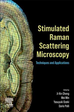
Stimulated Raman Scattering Microscopy: Techniques and Applications
Editat de Ji-Xin Cheng, Wei Min, Yasuyuki Ozeki, Dario Pollien Limba Engleză Paperback – 7 dec 2021
This rapidly growing field needs a comprehensive resource that brings together the current knowledge on the topic, and this book does just that. Researchers who need to know the requirements for all aspects of the instrumentation as well as the requirements of different imaging applications (such as different types of biological tissue) will benefit enormously from the examples of successful demonstrations of SRS imaging in the book.
Led by Editor-in-Chief Ji-Xin Cheng, a pioneer in coherent Raman scattering microscopy, the editorial team has brought together various experts on each aspect of SRS imaging from around the world to provide an authoritative guide to this increasingly important imaging technique. This book is a comprehensive reference for researchers, faculty, postdoctoral researchers, and engineers.
- Includes every aspect from theoretic reviews of SRS spectroscopy to innovations in instrumentation and current applications of SRS microscopy
- Provides copious visual elements that illustrate key information, such as SRS images of various biological samples and instrument diagrams and schematics
- Edited by leading experts of SRS microscopy, with each chapter written by experts in their given topics
Preț: 1030.51 lei
Preț vechi: 1668.36 lei
-38% Nou
Puncte Express: 1546
Preț estimativ în valută:
197.19€ • 206.40$ • 164.12£
197.19€ • 206.40$ • 164.12£
Carte tipărită la comandă
Livrare economică 25 martie-08 aprilie
Preluare comenzi: 021 569.72.76
Specificații
ISBN-13: 9780323851589
ISBN-10: 0323851584
Pagini: 610
Ilustrații: Approx. 300 illustrations
Dimensiuni: 216 x 276 mm
Greutate: 1.4 kg
Editura: ELSEVIER SCIENCE
ISBN-10: 0323851584
Pagini: 610
Ilustrații: Approx. 300 illustrations
Dimensiuni: 216 x 276 mm
Greutate: 1.4 kg
Editura: ELSEVIER SCIENCE
Public țintă
Primary: Researchers, faculty, postdoctoral researchers, and engineers interested in SRS technology/imaging; chemists and biomedical scientists in industrySecondary: This book can be used as a reference for a graduate level course
Cuprins
Preface
Sunney Xie
Part 1: Theory
1. Coherent Raman scattering processes
2. Sensitivity and noise in SRS microscopy
3. Stimulated Raman Scattering: ensembles to single molecules
Part 2: Advanced Instrumentation and Emerging Modalities
4. Hyperspectral SRS imaging via spectral focusing
5. Balanced detection SRS microscopy
6. Multiplex stimulated Raman Scattering microscopy via a tuned amplifier
7. Impulsive SRS microscopy
8. Multicolor SRS imaging with wavelength-tunable/switchable lasers
9. Pulse-shaping-based SRS spectral imaging and applications
10. Background-free stimulated Raman scattering imaging by manipulating photons in the spectral domain
11. Coherent Raman scattering microscopy for superresolution vibrational imaging: Principles, techniques, and implementations
12. Quantum-enhanced stimulated Raman scattering
13. Stimulated Raman excited fluorescence (SREF) microscopy: Combining the best of two worlds
14. Instrumentation and methodology for volumetric stimulated Raman scattering imaging
15. SRS flow and image cytometry
16. Widely and rapidly tunable fiber laser for high-speed multicolor SRS
17. Compact fiber lasers for stimulated Raman scattering microscopy
18. Synchronized time-lens source for coherent Raman scattering microscopy
Part 3: Vibrational Probes
19. Spontaneous Raman and SERS microscopy for Raman tag imaging
20. Stimulated Raman scattering imaging with small vibrational probes
21. Supermultiplexed vibrational imaging: From probe development to biomedical application
22. Raman beads for bio-imaging
23. Plasmon-enhanced stimulated Raman scattering microscopy
Part 4: Data Science
24. Converting hyperspectral SRS into chemical maps
25. Compressive Raman microspectroscopy
26. Denoise SRS images
Part 5: Applications to Life Sciences and Materials Science
27. Use of SRS microscopy for imaging drugs
28. Isotope-probed SRS (ip-SRS) imaging of metabolic dynamics in living organisms
29. Rapid determination of antimicrobial susceptibility by SRS single-cell metabolic imaging
30. Stimulated Raman scattering imaging of cancer metabolism: New avenue to precision medicine
31. Biomedical applications of SRS microscopy in functional genetics and genomics
32. Stimulated Raman voltage imaging for quantitative mapping of membrane potential
33. Neurodegenerative disease by SRS microscopy
34. Applications of stimulated Raman scattering (SRS) microscopy in materials science
35. Resolving molecular orientation by polarization-sensitive stimulated Raman scattering microscopy
Part 6: Miniaturization and Translation to Medicine
36. Stimulated Raman histology
37. Miniaturized handheld stimulated Raman scattering microscope
38. Intraoperative multimodal imaging
