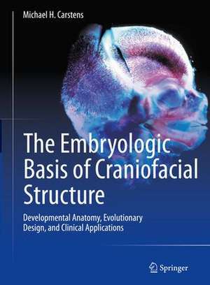
The Embryologic Basis of Craniofacial Structure: Developmental Anatomy, Evolutionary Design, and Clinical Applications
Editat de Michael H. Carstensen Limba Engleză Hardback – 28 sep 2023
The first section is meant for readers to gain a fundamental understanding of the development of craniofacial structures, from embryo onward, relying on the concepts of the Neuromeric Theory. The chapters in the first section of the book trace the development of the typical patient. The second section offers clinical examples of how the Neuromeric Theory can be used to repair or reconstruct various regions of the head and neck. Craniofacial clefts, including cleft lip and palate, ocular hypotelorism, anencephaly, craniosynostosis and more are detailed. Understanding the formation of the tissue structures involved in any given genetic deformation or anomaly enables the clinician to provide a more satisfying outcome for the patient, both structurally and aesthetically. New and current therapeutic options are explored and supported through original illustrations and photographs to aid in determining the best treatment for each individual patient.
Embryological Principles of Craniofacial Structure bridges the gap between introductory books on the basic anatomy of the head and neck and the detailed understanding required for corrective surgery of craniofacial defects.
Preț: 1540.95 lei
Preț vechi: 1630.14 lei
-5% Nou
Puncte Express: 2311
Preț estimativ în valută:
294.100€ • 303.39$ • 244.73£
294.100€ • 303.39$ • 244.73£
Carte indisponibilă temporar
Doresc să fiu notificat când acest titlu va fi disponibil:
Se trimite...
Preluare comenzi: 021 569.72.76
Specificații
ISBN-13: 9783031156359
ISBN-10: 3031156358
Pagini: 1723
Ilustrații: XLVIII, 1723 p. 1718 illus., 1387 illus. in color. With online files/update. In 2 volumes, not available separately.
Dimensiuni: 210 x 279 mm
Greutate: 6.19 kg
Ediția:1st ed. 2023
Editura: Springer International Publishing
Colecția Springer
Locul publicării:Cham, Switzerland
Cuprins
Chapter 1. Neuromeric Organization of the Head and Neck.- Chapter 2. Anatomy of Mesenchyme and the Pharyngeal Arches.- Chapter 3. Carnegie Staging System.- Chapter 4. Neurovascular Organization and Assembly of the Face.- Chapter 5. The Neuromeric System: Segmentation of the Neural Tube.- Chapter 6. Development of the Craniofacial Blood Supply – Cerebrovascular System.- Chapter 7. Vascular System, 2: Face, Orbit, and Meninges.- Chapter 8. Developmental Anatomy of the Craniofacial Bones.- Chapter 9. Neuromuscular Development: Motor Columns, Cranial Nerves, and Pharyngeal Arches.- Chapter 10. The Neck: Development and Evolution.- Chapter 11. Developmental Anatomy of Craniofacial Skin and Fascia.- Chapter 12. The Meninges.- Chapter 13. The Orbit.- Chapter 14. Pathologic Anatomy of the Hard Palate.- Chapter 15. Alveolar Extension Palatoplasty: The Role of Developmental Field Reassignment in the Prevention of Sequential Vascular Isolation and Growth Arrest.- Chapter 16. Pathologic Anatomy of the Soft Palate.- Chapter 17. Buccinator Interposition Palatoplasty: The Role of Developmental Field Reassignment in the Management of Velopharyngeal Insufficiency.- Chapter 18. Pathologic Anatomy of Nasolabial Clefts: Spectrum of the Microform Deformity and the Neuromeric Basis of Cleft Surgery.- Chapter 19. DFR Cheilorhinoplasty: The Role of Developmental Field Reassignment in the Management of Facial Asymmetry and the Airway in the Complete Cleft Deformity.- Chapter 20. Biologics in Craniofacial Reconstruction: Morphogens and Stem Cells.
Notă biografică
Michael H. Carstens, MD, FACS
Wake Forest Institute of Regenerative Medicine
Wake Forest University
Winston-Salem, North Carolina
USA
Dr. Michael Carstens received his A.B. in Latin American Studies with honors and Phi Beta Kappa from Stanford University. He subsequently completed a B.S. in chemistry with honors from Colorado State University. He received his M.D. from Stanford. He completed residency training in general surgery at Boston University and in plastic surgery at the University of Pittsburgh. He holds two fellowships in craniofacial surgery from the University of Pittsburgh and from the University of Southern California. He is currently working with the Wake Forest Institute of Regenerative Medicine at Wake Forest University and teaches in several medical schools around the world. His academic interests are craniofacial embryology, the developmental origin of stem cells and the surgical management of facial clefts.
Dr. Carstens speaks six languages; this makes him a well-respected ambassador for craniofacial surgery around the world. His career has been remarkable for social involvement. In 1990-1992 a consultancy for the World Health Organization brought Dr. Carstens to Nicaragua; he became a co-founder of the renowned APROQUEN foundation for burn care in that country. Currently he works for WFIRM and serve as scientific consultant in regenerative medicine for the Ministry of Health of Nicaragua where he continues doing research on developmental embryology and clinical applications of stromal vascular fraction (SVF) cells for complex wounds and chronic inflammatory states.
Textul de pe ultima copertă
Focusing on the anatomy of the head and neck, this book begins at the cellular level of development, detailing bone, muscle, blood supply, and innervation along the way. It illustrates the origin of each tissue structure to aid in making prognoses beyond the surface deformation, offering typical issues seen in the craniofacial region, for example. Written by a pediatric Craniofacial plastic surgeon and intended for clinicians and residents in the areas of plastic surgery, ENT, maxillofacial surgery, and orthodontistry, this book is the first of its kind to focus so intently on evolution of the craniofacial structure. It is neatly broken up into two distinct sections.
The first section is meant for readers to gain a fundamental understanding of the development of craniofacial structures, from embryo onward, relying on the concepts of the Neuromeric Theory. The chapters in the first section of the book trace the development of the typical patient. The second section offers clinical examples of how the Neuromeric Theory can be used to repair or reconstruct various regions of the head and neck. Craniofacial clefts, including cleft lip and palate, ocular hypotelorism, anencephaly, craniosynostosis and more are detailed. Understanding the formation of the tissue structures involved in any given genetic deformation or anomaly enables the clinician to provide a more satisfying outcome for the patient, both structurally and aesthetically. New and current therapeutic options are explored and supported through original illustrations and photographs to aid in determining the best treatment for each individual patient.
Embryological Principles of Craniofacial Structure bridges the gap between introductory books on the basic anatomy of the head and neck and the detailed understanding required for corrective surgery of craniofacial defects.
Embryological Principles of Craniofacial Structure bridges the gap between introductory books on the basic anatomy of the head and neck and the detailed understanding required for corrective surgery of craniofacial defects.
Caracteristici
Uses the Neuromeric Theory to explain the basis of treating genetic anomalies
Provides an interdisciplinary understanding of a notoriously difficult to understand region of the body
Features original illustrations and photographs, animations of techniques, and video of procedures
Provides an interdisciplinary understanding of a notoriously difficult to understand region of the body
Features original illustrations and photographs, animations of techniques, and video of procedures
