The Equine Distal Limb: An Atlas of Clinical Anatomy and Comparative Imaging
Autor Jean-Marie Denoixen Limba Engleză Hardback – 11 iul 2000
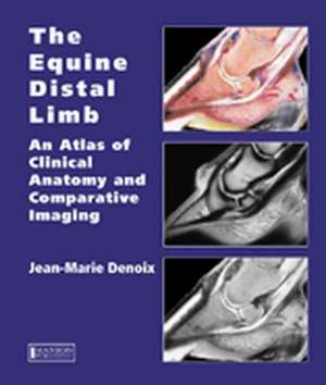
This book provides a unique contribution and is an invaluable reference text. The image quality is extraordinarily high and the multiple views of each area of the distal limb provide an extremely detailed evaluation. Each part opens with a characteristically superb anatomical drawing by the author. Each spread of pages deals with a single dissection viewed by means of colour photographs, labelled black and white equivalents, plus x-rays, ultrasound and MRI scans as required. The book is designed for maximum clarity using a generous page size. The atlas is essential for anybody involved in detailed anatomical study, complex lameness evaluation or advanced imaging techniques.
Preț: 1605.71 lei
Preț vechi: 1764.51 lei
-9% Nou
Puncte Express: 2409
Preț estimativ în valută:
307.29€ • 317.02$ • 260.08£
307.29€ • 317.02$ • 260.08£
Carte disponibilă
Livrare economică 12-26 februarie
Livrare express 28 ianuarie-01 februarie pentru 100.80 lei
Preluare comenzi: 021 569.72.76
Specificații
ISBN-13: 9781840760019
ISBN-10: 184076001X
Pagini: 400
Ilustrații: 1212 colour illustrations
Dimensiuni: 250 x 291 x 25 mm
Greutate: 2.46 kg
Ediția:1
Editura: CRC Press
Colecția CRC Press
Locul publicării:Chichester, United Kingdom
ISBN-10: 184076001X
Pagini: 400
Ilustrații: 1212 colour illustrations
Dimensiuni: 250 x 291 x 25 mm
Greutate: 2.46 kg
Ediția:1
Editura: CRC Press
Colecția CRC Press
Locul publicării:Chichester, United Kingdom
Public țintă
Professional Practice & DevelopmentCuprins
The Equine Foot: Dissections of the equine foot. Sagittal and parasagittal sections of the equine foot. Transverse sections of the Equine Foot. Frontal sections of the equine foot.
The Equine Pastern: Dissections of the equine pastern. Sagittal and parasagittal sections of the equine pastern. Transverse sections of the equine pastern. Frontal sections of the equine pastern.
The Equine Fetlock: Dissections of the equine fetlock. Sagittal and parasagittal sections of the equine fetlock. Transverse sections of the equine fetlock. Frontal sections of the equine fetlock.
Descriere
Jean-Marie Denoix is the world’s leading equine musculoskeletal system anatomist and has become one of the foremost equine diagnostic ultrasonographers. There is therefore nobody better to compile a reference atlas of the clinical anatomy of the foot, pastern and fetlock, correlated with images obtained by radiography, diagnostic ultrasonography and magnetic resonance imaging. Advanced imaging techniques require in depth knowledge of anatomy for accurate interpretation and especially when using magnetic resonance imaging this must be a 3-dimensional concept of anatomy.


