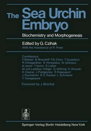
The Sea Urchin Embryo: Biochemistry and Morphogenesis
Editat de G. Czihak Cuvânt înainte de J. Brachet Contribuţii de R. Peter, F. Baltzeren Limba Engleză Paperback – 6 dec 2011
Preț: 666.41 lei
Preț vechi: 784.01 lei
-15% Nou
Puncte Express: 1000
Preț estimativ în valută:
127.51€ • 133.50$ • 105.51£
127.51€ • 133.50$ • 105.51£
Carte tipărită la comandă
Livrare economică 07-21 aprilie
Preluare comenzi: 021 569.72.76
Specificații
ISBN-13: 9783642659669
ISBN-10: 3642659667
Pagini: 728
Ilustrații: XX, 702 p.
Dimensiuni: 170 x 244 x 38 mm
Greutate: 1.14 kg
Ediția:Softcover reprint of the original 1st ed. 1975
Editura: Springer Berlin, Heidelberg
Colecția Springer
Locul publicării:Berlin, Heidelberg, Germany
ISBN-10: 3642659667
Pagini: 728
Ilustrații: XX, 702 p.
Dimensiuni: 170 x 244 x 38 mm
Greutate: 1.14 kg
Ediția:Softcover reprint of the original 1st ed. 1975
Editura: Springer Berlin, Heidelberg
Colecția Springer
Locul publicării:Berlin, Heidelberg, Germany
Public țintă
ResearchDescriere
Sea urchin eggs are objects of wonder for the student who sees them for the first time under the microscope. The formation of the fertil ization membrane after insemination, the beauty of mitotic cleavage, the elegant swimming of embryos, remain an esthetic pleasure even for the eyes of seasoned investigators. But sea urchin eggs have other, more practical, advantages: they lend themselves to surgical operation without difficulty and they heal perfectly; they can be obtained in very large amounts and represent thus an extremely favorable material for biochemists and molecular embryologists. It is not surprising that, in view of these exceptional advantages, sea urchin eggs have attracted the interest of innumerable biologists since O. HERTWIG discovered the fusion of the pronuclei (amphimixy), in Paracentrotus lividus, almost a century ago. The purpose of the present book is to present, in a complete and orderly fashion, the enormous amount of information which has been gathered, in the course of a hun dred years of sea urchin embryology. JOSEPH NEEDHAM, in 1930, was still able to present all that was known, at that time, on the biochemistry of all possible species of developing eggs and embryos in his famous "Chemical Embryology" (Cambridge University Press) . It would no longer be possible for one man to write a modern version of what was a "Bible" for the young embryologists of forty years ago.
Cuprins
1. Introductory History.- 2A. Care and Handling of Sea Urchin Eggs, Embryos, and Adults (Principally North American Species).- 2A.1 Collecting and Keeping.- 2A.2 Spawning Season.- 2A.3 Determining Sex.- 2A.4 Obtaining Gametes.- 2A.5 Preparing Eggs.- 2A.6 Counting Eggs.- 2A.7 Storing Gametes.- 2A.8 Agents Affecting Development.- 2A.9 Choosing Eggs.- 2A.10 Fertilization.- 2A.11 Removing Extracellular Structures.- 2A.12 Normal Development.- 2B. Handling Japanese Sea Urchins and Their Embryos.- 2B.1 General Handling.- 2B.2 Echinoids as Experimental Materials.- 2B.3 Maintenance.- 2B.4 Sexual Dimorphism.- 2B.5 Artificial Spawning.- 2B.6 Gametes.- 2B.7 Artificial Fertilization.- 2B.8 Development and Growth.- 2C. Maturation Periods of European Sea Urchin Species.- 3. Gametogenesis.- 3.1 Gonad Biology.- 3.2 Germ Cells.- 3.3 Immature Gonads.- 3.4 Spermatogenesis.- 3.5 Oogenesis.- 3.6 Conclusions.- 4. Fertilization.- 4.1 Activity of Spermatozoa.- 4.2 Acrosome Reaction.- 4.3 Spermatozoon Penetration.- 4.4 Partial Fertilization.- 4.5 Egg Activation by Sperm Injection.- 4.6 Sperm Role in Egg Activation.- 4.7 Egg Cortical Response.- 4.8 Narcotized Egg Responses.- 4.9 Refertilization and Polyspermy.- 4.10 Conclusions.- 5. Parthenogenetic Activation and Development.- 5.1 Ways of Inducing Parthenogenetic Development.- 5.2 How Parthenogenetic Activation Works.- 5.3 Conclusions.- 6. Cytology of Parthenogenetically Activated Sea Urchin Eggs.- 6.1 Monaster Formation.- 6.2 Cytaster Formation.- 6.3 Karyogene Spindle Formation.- 6.4 Haploid Mitosis.- 6.5 Combinations.- 7. Normal Development to Metamorphosis.- 7.1 Cleavage Stage.- 7.2 The Blastula before Hatching.- 7.3 The Blastula after Hatching.- 7.4 Gastrula Stage.- 7.5 Prism and Pluteus Stages.- 7.6 Late Larval Stage and Metamorphosis.- 8. Cellular Behavior and Cytochemistry in Early Stages of Development.- 8.1 The Sea-Urchin Embryo in Studies of Morphogenetic Cell Behavior.- 8.2 Morphogenetic Cell Behavior Analysis.- 8.3 Cleavage and Blastula Formation.- 8.4 Morphogenetic Cell Behavior in the Ectoderm.- 8.5 Morphogenetic Cell Behavior in the Mesendodermal and Mesenchymal Region.- 8.6 Cytologic Aspects of Filopodal Attachments.- 8.7 Morphogenetic Cell Behavior during Further Development of the Mesendoderm.- 8.8 Biochemical Differentiation of the Blastula and Gastrula.- 8.9 Biochemical Activity and Morphogenetic Movement.- 9. Egg and Embryo Ultrastructure.- 9.1 The Mature Egg.- 9.2 The Mature Spermatozoon.- 9.3 Fertilization.- 9.4 Pronuclear Development and Fusion.- 9.5 Mitosis.- 9.6 From 2-Cell Stage to Gastrula.- 10. The Biophysics of Cleavage and Cleavage of Geometrically Altered Cells.- 10.1 General.- 10.2 Cleavage Theories.- 10.3 Biophysics of Cleavage.- 10.4 Cleavage Pattern Alteration.- 10.5 Summary.- 11. Centrifugation and Alteration of Cleavage Pattern.- 11.1 Techniques.- 11.2 Morphological Changes.- 11.3 Electron Microscope Studies.- 11.4 Developmental Patterns.- 11.5 Cleavage Patterns and Micromere Formation.- 11.6 Development of the Dorso-Ventral Axis.- 11.7 Respiration.- 11.8 Biochemistry.- 12. Irradiation, Chemical Treatment, and Cleavage-Cycle and Cleavage-Pattern Alteration.- 12.1 Stimulation of Premature Division.- 12.2 Division Delay.- 12.3 Division Abnormalities.- 12.4 Early Post-Fertilization Changes.- 12.5 The General Cell Cycle.- 12.6 DNA Synthesis.- 12.7 RNA Synthesis.- 12.8 Protein Synthesis.- 12.9 Pronuclear Fusion.- 12.10 Duplication and Separation of Centrioles.- 12.11 Chromosomal Condensation and Nuclear Membrane Breakdown.- 12.12 Metaphase, Anaphase, and Telophase.- 13. Isolation and Transplantation Experiments.- 13.1 Methods.- 13.2 Determination of Cleavage.- 13.3 Determination along the Egg Axis.- 13.4 Dorsoventral Axis Determination.- 13.5 Interactions between Species.- 13.6 Fragments Used for Physiological Tests.- 14. Blastomere Reaggregation.- 15. Morphology and Biochemistry of Diploid and Androgenetic Haploid (Merogonic) Hybrids.- 15.1 Classification.- 15.2 Hybrid Fertilization Techniques.- 15.3 Early Development of Parental Species.- 15.4 Developmental Capacity and Morphologic Characteristics of Hybrids.- 15.5 Biochemical Properties of Hybrids.- 15.6 Conclusions.- 16. Animalization and Vegetalization.- 16.1 Introduction.- 16.2 Definitions.- 16.3 Experimental Methods.- 16.4 Vegetalizing Agents.- 16.5 Animalizing Agents.- 16.6 Research on the Metabolism of Animalized and Vegetalized Larvae.- 16.7 Biophysical Aspects.- 16.8 Concluding Remarks.- Addendum.- 17. Respiration and Energy Metabolism.- 17.1 Overall Changes in Respiration during Development.- 17.2 Increased Respiration after Fertilization.- 17.3 Respiratory Change after Fertilization.- 17.4 Respiratory Increase during Development; Energy Metabolism.- 17.5 High-Energy Phosphate Compounds and Their Metabolic Activity.- 18. Carbohydrate Metabolism and Related Enzymes.- 18.1 Glycolysis and the Pentose Phosphate Shunt.- 18.2 Pyruvate Metabolism and the Tricarboxylic Acid (KREBS-) Cycle.- 18.3 Terminal Electron Transport System.- 19. Lipids.- 19.1 Lipids in General.- 19.2 Phospholipids.- 19.3 Glycolipids.- 19.4 Cholesterol.- 19.5 Carotenoids.- 19.6 Lipid Metabolism.- 20. DNA in Gametes and Early Development.- 20.1 DNA in Gametes.- 20.2 DNA Synthesis during Development.- 20.3 Enzymes.- 21. Integrating Factors.- 21.1 Organization of Egg Cytoplasm.- 21.2 Differentiation along the Egg Axis.- 21.3 Integration of DNA and Nuclear Protein.- 21.4 Differentiation along the Dorsoventral Axis.- 21.5 Summary.
